. 2010 Jul 16;285(29):21922-33.
doi: 10.1074/jbc.M110.132027. Epub 2010 May 12.

The eukaryotic initiation factor (eIF) 4G HEAT domain promotes translation re-initiation in yeast both dependent on and independent of eIF4A mRNA helicase

Affiliations

The eukaryotic initiation factor (eIF) 4G HEAT domain promotes translation re-initiation in yeast both dependent on and independent of eIF4A mRNA helicase

Ryosuke Watanabe et al. J Biol Chem. .

Abstract

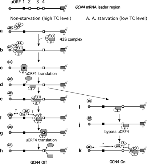
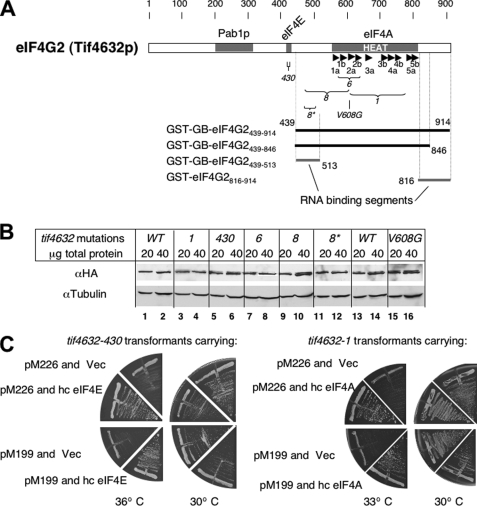
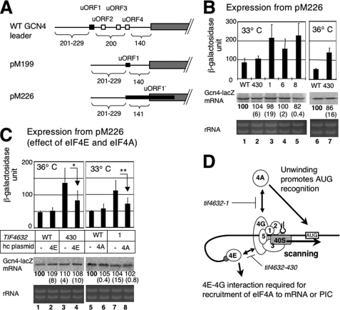
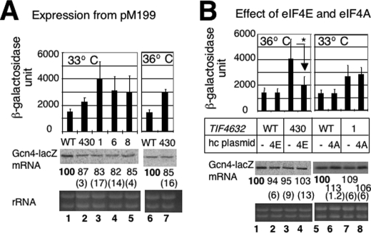
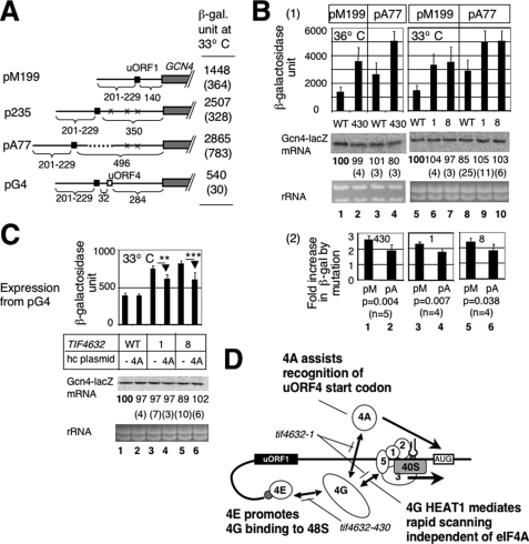
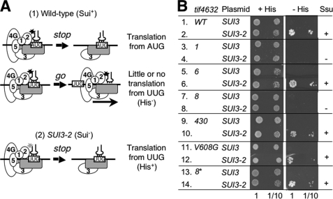
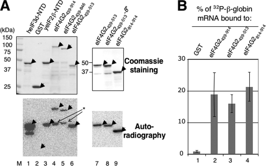
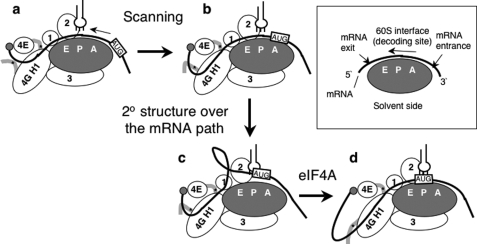
Translation re-initiation provides the molecular basis for translational control of mammalian ATF4 and yeast GCN4 mediated by short upstream open reading (uORFs) in response to eIF2 phosphorylation. eIF4G is the major adaptor subunit of eIF4F that binds the cap-binding subunit eIF4E and the mRNA helicase eIF4A and is also required for re-initiation in mammals. Here we show that the yeast eIF4G2 mutations altering eIF4E- and eIF4A-binding sites increase re-initiation at GCN4 and impair recognition of the start codons of uORF1 or uORF4 located after uORF1. The increase in re-initiation at GCN4 was partially suppressed by increasing the distance between uORF1 and GCN4, suggesting that the mutations decrease the migration rate of the scanning ribosome in the GCN4 leader. Interestingly, eIF4E overexpression suppressed both the phenotypes caused by the mutation altering eIF4E-binding site. Thus, eIF4F is required for accurate AUG selection and re-initiation also in yeast, and the eIF4G interaction with the mRNA-cap appears to promote eIF4F re-acquisition by the re-initiating 40 S subunit. However, eIF4A overexpression suppressed the impaired AUG recognition but not the increase in re-initiation caused by the mutations altering eIF4A-binding site. These results not only provide evidence that mRNA unwinding by eIF4A stimulates start codon recognition, but also suggest that the eIF4A-binding site on eIF4G made of the HEAT domain stimulates the ribosomal scanning independent of eIF4A. Based on the RNA-binding activities identified within the unstructured segments flanking the eIF4G2 HEAT domain, we discuss the role of the HEAT domain in scanning beyond loading eIF4A onto the pre-initiation complex.


Figures

FIGURE 1.
FIGURE 1.
Model for GCN4 translational control. Lines indicate the GCN4 mRNA leader with the gray boxes to the right (followed by diagonal hashed lines) representing the GCN4 coding region and the gray circle to the left representing the 5′ cap. Of the four uORFs, shown on top, two (uORFs 1 and 4) have been shown to be necessary and sufficient for regulation of GCN4 expression, and are depicted as filled and open squares, respectively, on the second line (panel a) and below (panels b–k). The figure illustrates the ribosome movement on the leader region with the focus on its association with eIF4G (4G), eIF4E (4E), eIF4A (4A), eIF1 (1), eIF3 (3), eIF5 (5), and eIF2 TC (T). eIF4E is associated with the cap loosely but strongly enough to protect it from decapping (dotted line between the cap and eIF4E). The cap interaction with eIF4E is enhanced by eIF4E binding to eIF4G (direct contact of the cap with eIF4E in panels a, b, e, f, and k). The 40 S and 60 S subunits are drawn as a gray round rectangle and gray oval, respectively. Under non-starvation conditions (left column), the preinitiation complex scans for and translates uORF1. After uORF1 translation, eIF3 bound to post-uORF1 termination 40 S prevents mRNA dissociation from the 40 S. As a consequence, a population of 40 S subunits remains associated with the mRNA and resumes scanning after reacquiring TC and other eIFs (panels e and f). Subsequent translation of uORF4 dissociates the ribosome, shutting off GCN4 translation (panels g and h). Under amino acid starvation conditions (right column), Gcn2p kinase is activated and phosphorylates eIF2. This phosphorylation renders eIF2 into a competitive inhibitor of GDP/GTP exchange activity (catalyzed by eIF2B; not shown), thereby reducing the level of eIF2/GTP and hence TC levels. uORF4 is bypassed due to low TC levels (panels i and j). TC and eIF4G can be re-acquired during scanning of the uORF4-GCN4 interval, resulting in translation of GCN4 (panel k). “?” in the figure indicates the uncertainty of the interaction of eIF4G with the cap-bound eIF4E, and the 48 S PIC migrating after uORF1 translation, which are addressed in this study.
FIGURE 2.
FIGURE 2.
eIF4G mutations used in this study. A, primary structure of eIF4G2 (Tif4632p) with defined binding sites for Pab1p, eIF4E, and eIF4A (gray boxes). Arrowheads denote α-helices numbered according to the structure of mammalian eIF4GII HEAT1 (17). Brackets indicate the regions of tif4632 mutations used in this study (see Table 3 for specific amino acid changes). Thick lines to the bottom represent the portion of eIF4G2 polypeptide carried by the four GST fusion constructs used here and indicated to the left (Table 1). Numbers indicate the eIF4G2 residues at the boundary of each construct. Thick gray lines indicate the RNA binding segments identified in this study (Fig. 7). B, immunoblot indicating equal abundance of HA-eIF4G2 mutants expressed from strains used in this study. Antibodies used are indicated to the left of each gel. Anti-HA detects HA-eIF4G2 and its mutants. Anti-tubulin is used as a loading control. Total protein amounts of cell extracts used are listed under mutations. Strain used are YAS1955 (WT), KAY108 (tif4632-1), KAY109 (tif4632-430), KAY110 (tif4632-6), KAY111 (tif4632-8), KAY872 (tif4632-8*), KAY220 (his4 WT), and KAY910 (his4 tif4632-V608G). C, plate assays demonstrating Ts phenotypes of tif4632-430 (KAY109, left panels) and tif4632-1 (KAY108, right panels) transformants carrying GCN4-lacZ reporter plasmids (pM226 and pM199 used in Figs. 3 and 4, respectively) and their suppression by YEpA-TIF45 (hc eIF4E) and YEpA-TIF2 (hc eIF4A), respectively (Vec, an empty vector). Transformants carrying indicated plasmids were streaked out on plates made of synthetic defined medium and incubated at temperature listed at the bottom for 3 days.
FIGURE 3.
FIGURE 3.
Effect of tif4632 mutations on GCN4-lacZ expression from pM226. A, the primary structure of GCN4 leader region, either wild-type (top) or one found in pM199 (middle) or pM226 (bottom). Filled box, uORF1, the positive regulatory element. Open boxes, uORF2–4, the negative regulatory elements. Gray box, GCN4 ORF. Numbers under the schematic indicate the distance in nucleotides between 5′-end of the transcript and uORF1 (numbers to the left) and that between major elements of the leader region. B, transformants of YAS1955 (WT) and its tif4632-1 (1), -6 (6), -8 (8), and -430 (430) derivatives (Table 3) carrying pM226 were grown at indicate temperatures in synthetic complete medium lacking uracil (SC-ura) and assayed for β-galactosidase activity (24). Graphs to the top indicate average values of the β-galactosidase activity units with bars denoting S.D. (see “Materials and Methods” for statistics). Middle and bottom gels: 40 μg of total RNA isolated from the same transformants grown under the same conditions was resolved by agarose gel electrophoresis and stained by ethidium bromide (bottom gel), followed by Northern blotting with 32P-lacZ probe (middle gel). Values under the middle gel indicate the relative levels of GCN4-lacZ mRNA determined by the band 32P intensity, averaged from two independent experiments. Values in parentheses are S.D. from these experiments. C, double transformants of yeast strains as defined in B carrying pM226 and a hc ADE2 plasmid carrying eIF4E (4E), eIF4A (4A) (Table 1) or an empty vector (−) grown in SC-ura-ade were assayed for β-galactosidase activity and mRNA levels, and the data are presented as described in B. p values for differences indicated by arrows are 0.016 (n = 5) for * and 0.015 (n = 8) for **. D, model of the eIF interactions in the pre-initiation complex during the process of scanning for the uORF1 start codon (box labeled AUG). Thick line indicates mRNA, with gray circle representing 5′ m7G cap. Empty circles labeled with numbers denote relevant eIFs. The plug touching eIF2 is Met-tRNAiMet with anticodon. Stopped bars indicate interactions (arrows) impaired by tif4632 mutations.
FIGURE 4.
FIGURE 4.
Effect of tif4632 mutations on GCN4-lacZ expression from pM199. Transformants of YAS1955 (WT) or its tif4632 derivatives (Table 3) carrying pM199 in the presence (B) or absence (A) of additional plasmid overexpressing eIF4A (4A) and eIF4E (4E) were assayed for β-galactosidase and Northern blotting, and the data were presented as described in Fig. 3B. In B, p values for difference indicated by * is 0.007 (n = 8).
FIGURE 5.
FIGURE 5.
Effect of tif4632 mutations on GCN4-lacZ expression from pA77 and pG4. A, modified GCN4 leader structures for the re-initiation reporter constructs used in this study, drawn with symbols defined in Fig. 3A. X indicates the presence of a start codon mutation in the corresponding uORF. Dotted line denotes a foreign sequence lacking start or stop codons. The table to the right summarizes β-galactosidase activity from YAS1955 transformants carrying the plasmids grown in SC-ura at 33 °C with S.D. in parenthesis. B and C, transformants of YAS1955 (WT) or its tif4632 derivatives (Table 3) carrying the indicated GCN4-lacZ plasmid in the presence (C) or absence (B and C) of an additional plasmid overexpressing eIF4A (4A) were assayed for β-galactosidase activity and mRNA levels; the data are presented as described in Fig. 3B. In B, panel 2 shows the ratio of β-galactosidase activity from pM199 (pM) or pA77 (pA) in the indicated mutant to that in wild type, based on the data in panel 1. p values for differences between the ratio with pM199 and one with pA77 are indicated below the graph. In C, p values for differences indicated by arrows are <0.001 (n = 6) for ** and 0.002 (n = 6) for ***. D, model of the eIF interactions in the pre-initiation complex during the process of scanning for a start codon (box labeled AUG) after uORF1 (filled box) translation. Symbols are as defined in Fig. 3D.
FIGURE 6.
FIGURE 6.
tif4632 mutations suppress relaxed start codon selection caused by an eIF2β mutation (SUI3-2). A, model of Sui phenotypes. A, panel 1, wild-type PIC fully loaded with different eIFs (circles with a number) can efficiently distinguish between AUG and UUG codons (boxed) on the mRNA (horizontal line), in part due to tight interaction (asterisk) between eIF2 and tRNAiMet (plug). If it encounters AUG, the PIC responds (stop) and releases eIFs to bind the 60 S, but if it encounters UUG, the PIC does not respond and resumes scanning (go). A, panel 2, when SUI3-2 impairs eIF2 interaction with tRNAiMet (light gray asterisk), the PIC encountering UUG erroneously responds (stop) and starts translation (Sui). B, 5 μl of 0.15 A600 unit cultures of transformants of KAY220 (his4-303) and its tif4632 mutant derivatives carrying YDpU-SUI3 (SUI3) or YDpU-SUI3-2 (SUI3-2) and their 10-fold dilution were spotted onto SC-ura (+His) and SC-ura-his (−His) plates and incubated at 30 °C for 3 and 7 days, respectively.
FIGURE 7.
FIGURE 7.
Determination of RNA binding activity carried by the interdomain segments of yeast eIF4G2. A, Northwestern blot experiment. Recombinant eIF4G2 (lanes 4–8) or control (lanes 1–3) proteins indicated across the top are immobilized on a nitrocellulose membrane after SDS-PAGE and incubated with 32P-β-globin mRNA, as described (27). After washing, the autoradiography was taken with a PhosphorImager (bottom panels). As a control, the same amount of proteins used in this experiment was subjected for SDS-PAGE and stained with Coomassie Blue (top gels). Arrowheads indicate the expected location of the full-length products. The asterisk indicates the RNA-binding activity of a proteolytically cleaved product, derived from GST-GB-eIF4G2439–914 or GST-GB-eIF4G2439–846. M, size standards. eIF4G2 proteins used here are described in Fig. 2A and were derived from pGEX-4G2 plasmids in Table 1. heIF3d-NTD and yeIF2β-NTD, which are fused to GST, were expressed from pGEX-p66ΔE (28) and pKA883,4 respectively. B, GST-eIF4G2 proteins or GST alone indicated at the top were allowed to bind 32P-β-globin mRNA (∼20,000 cpm) and the percentage of the RNA co-precipitated by a glutathione resin (average from three independent experiments) was determined by scintillation counting and presented with bars indicating S.D.
FIGURE 8.
FIGURE 8.
Model depicting the roles of eIF4G HEAT domain 1 (4G H1) and eIF4A in scanning and AUG selection. The scanning starts with the 40 S subunit (gray oval) bound to the mRNA (thick line) near the 5′-cap (gray circle) (a) and continues as the mRNA migrates over the path on the 40 S subunit (b). The model hypothesizes that a secondary structure develops as mRNA migrates over the path and thereby prevents correct matching of tRNAiMet (plug) anticodon to start codon (boxed and labeled AUG) (c). eIF4A resolves the structure and promotes AUG recognition (d). In addition to eIF4A, RNA binding to eIF4G HEAT1 and/or its surrounding peptides (shown by thick gray line with asterisk indicating the RNA-binding site) prevents mRNA refolding, thereby promoting scanning and AUG recognition. The orientation of eIF4G HEAT1 relative to the 40 S is based on the speculative model proposed recently (4). eIF4G HEAT1 binding to eIF1 (circle labeled 1) located near the E-site (9, 43) is proposed to contribute to maintaining a scanning-competent conformation (18). The box describes the 40 S subunit structure with A-, P-, and E-site and the orientation of bound mRNA.

Similar articles

Cited by

References

    1. Hinnebusch A. G., Dever T. E., Asano K. (2007) in Translational Control in Biology and Medicine (Mathews M. B., Sonenberg N., Hershey J. W. eds) Cold Spring Harbor Laboratory Press, Cold Spring Harbor, NY: 225–268
    1. Pestova T. V., Lorsch J. R., Hellen C. U. (2007) in Translational Control in Biology and Medicine (Mathews M. B., Sonenberg N., Hershey J. W. eds) Cold Spring Harbor Laboratory Press, Cold Spring Harbor, NY: 87–128
    1. Pisarev A. V., Kolupaeva V. G., Yusupov M. M., Hellen C. U., Pestova T. V. (2008) EMBO J. 27, 1609–1621 - PMC - PubMed
    1. Marintchev A., Edmonds K. A., Maritcheva B., Hendrickson E., Oberer M., Suzuki C., Herdy B., Sonenberg N., Wagner G. (2009) Cell 136, 447–460 - PMC - PubMed
    1. Asano K., Sachs M. S. (2007) Genes Dev. 21, 1280–1287 - PubMed

Publication types

MeSH terms

LinkOut - more resources