. 2010 Jul 23;285(30):23019-31.
doi: 10.1074/jbc.M110.100602. Epub 2010 May 12.

Molecular and functional basis for the scaffolding role of the p50/dynamitin subunit of the microtubule-associated dynactin complex

Affiliations

Molecular and functional basis for the scaffolding role of the p50/dynamitin subunit of the microtubule-associated dynactin complex

Guillaume Jacquot et al. J Biol Chem. .

Abstract

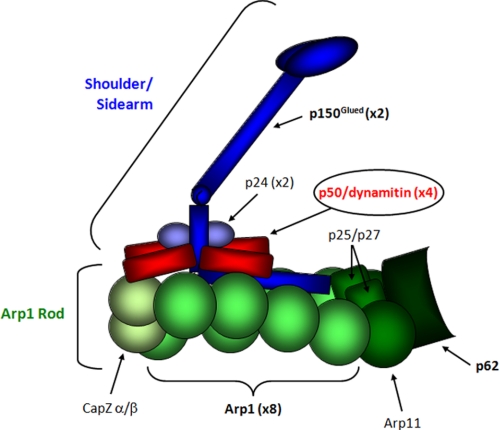
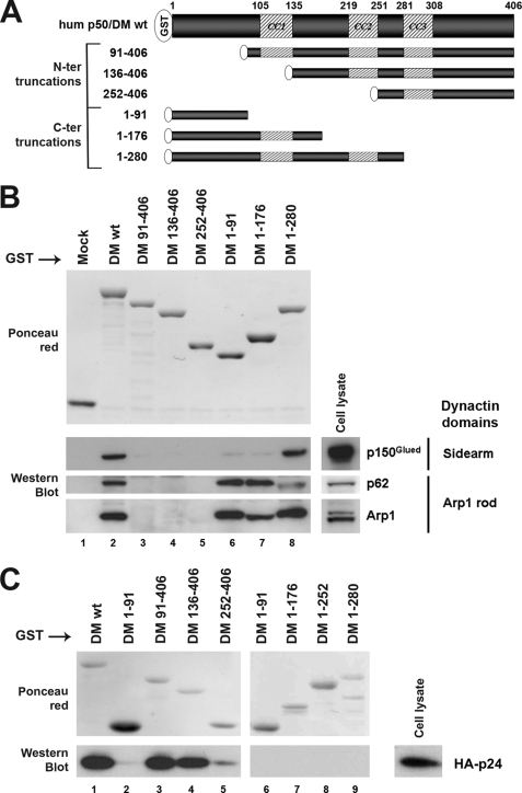
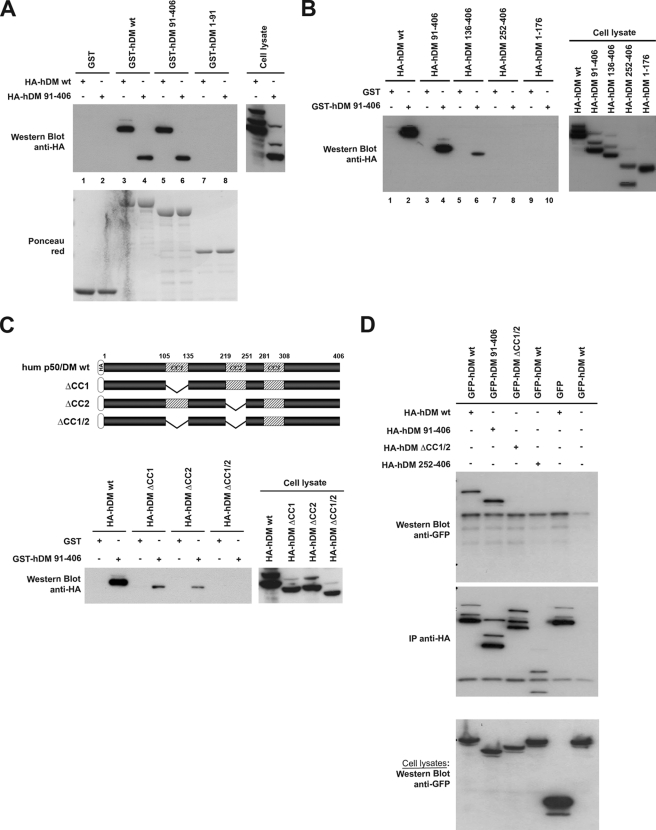
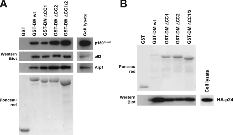
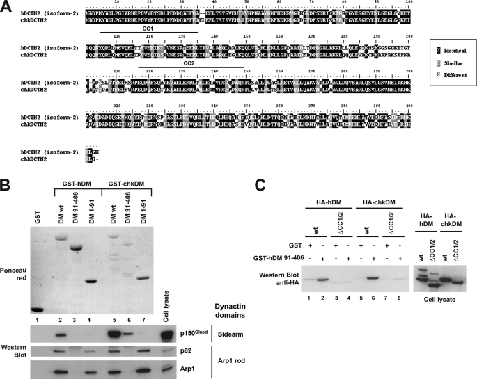
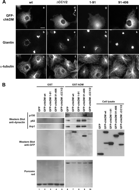
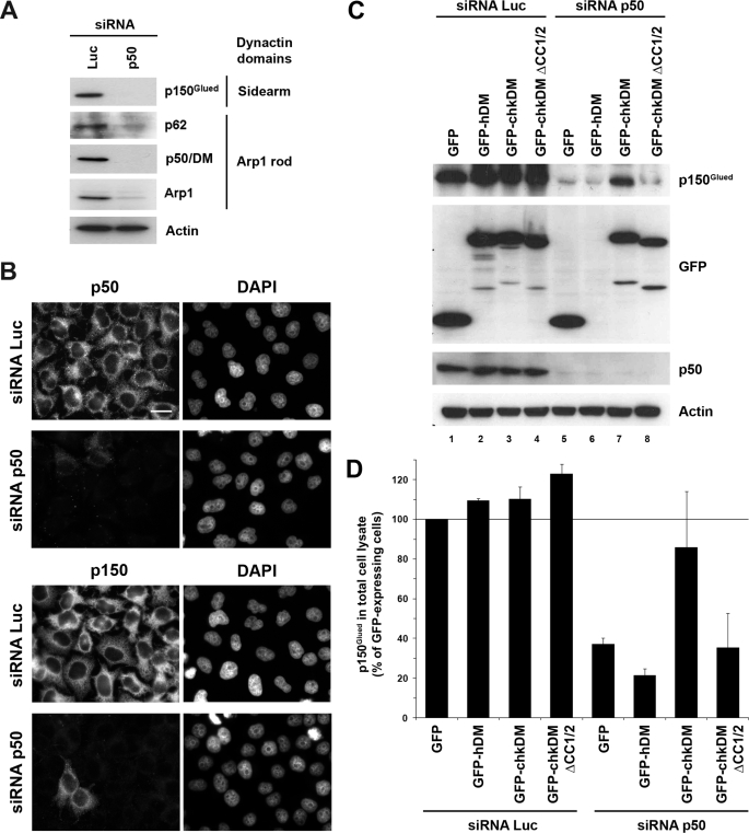
p50/dynamitin (DM) is a major subunit of the microtubule-associated dynactin complex that is required for stabilization and attachment of its two distinct structural domains, namely the Arp1 rod and the shoulder/sidearm. Here, we define the determinants of p50/DM required for self-oligomerization of the protein and for interactions with other subunits of the dynactin complex. Whereas the N-terminal 1-91-amino acid region of the protein is required and sufficient for binding to the Arp1 rod, additional determinants contained within the first half of the protein are required for optimal recruitment of the p150(Glued) subunit of the shoulder/sidearm. Overexpression experiments confirmed that the N-terminal 1-91-amino acid region of p50/DM is critical for dynactin functionality, because this fragment acts as a dominant negative to inhibit both dynein-dependent and -independent functions of the complex. Furthermore, the first two predicted coiled-coil motifs of p50/DM contain determinants that mediate self-association of the protein. Interestingly, p50/DM self-association does not contribute to p50/DM-induced disruption of the dynactin complex, but most likely participates in the stabilization of the complex.


Figures

FIGURE 1.
FIGURE 1.
Schematic representation of the overall structure of the dynactin complex. This schema illustrates the location and approximate structural features of individual subunits based on ultrastructural and biochemical characterization; it was adapted from Ref. .
FIGURE 2.
FIGURE 2.
Impact of human and chicken p50/DM overexpression on dynactin functions. A, COS7 cells expressing GFP, GFP-hDM, or GFP-chkDM (panels a, d, and g) were stained with an anti-Giantin pAb (panels b, e, and h) and an anti-α-tubulin mAb (panels c, f, and i). Arrows and arrowheads on panels d–f and g–i show transfected cells expressing high and low levels of the GFP-hDM fusions, respectively. Scale bar, 10 μm. B, Golgi distribution (black bars) and microtubule (MT) organization (gray bars) were quantified over 100 cells expressing GFP (mock), GFP-hDM (hDM), or GFP-chkDM (chkDM). Results are expressed as the percentage of transfected cells showing normal Golgi distribution, either concentrated in the perinuclear region (see nontransfected cells) or showing only slight dispersal (see arrowhead in panel e), and a radial organization of microtubules anchored at the centrosome. Values are the means of three independent experiments; error bars represent 1 standard deviation from the mean.
FIGURE 3.
FIGURE 3.
Characterization of the p50/DM determinants mediating interactions with other dynactin components. A, schematic representation of the human p50/DM deletion mutants fused to GST. Hatched segments represent the three putative coiled-coil motifs (CC1, CC2, and CC3); amino acids are numbered according to Yue et al. (36). wt, wild type; N-ter, N-terminal; C-ter, C-terminal. B, HeLa cell lysates (lower right panels) were incubated with equal amounts of wild type or deleted GST-hDM fusions (upper panel, Ponceau red) immobilized on GSH-Sepharose beads. Bound proteins were analyzed by Western blot with anti-p150, -p62, and -Arp1 antibodies. C, lysates from HeLa cells expressing HA-tagged p24 (lower right panel) were incubated with equal amounts of wild type or deleted GST-hDM fusions (upper panels, Ponceau red) immobilized on GSH-Sepharose beads. Bound proteins were analyzed as in B with anti-HA mAb.
FIGURE 4.
FIGURE 4.
Characterization of the determinants involved in p50/DM self-association. A, lysates from 293T cells expressing HA-tagged forms of either wild type hDM or the hDM-(91–406) mutant (upper right panel) were incubated with equal amounts of GST, GST-hDM wild type (wt), GST-hDM-(91–406), or GST-hDM-(1–91) (lower panel, Ponceau red) immobilized on GSH-Sepharose beads. Bound proteins were analyzed by Western blot with anti-HA mAb (upper left panel). B, lysates from 293T cells expressing wild type or deleted HA-tagged hDM (right panel) were incubated with equal amounts of GST or GST-hDM-(91–406) immobilized on GSH-Sepharose beads. Bound proteins were analyzed as indicated in A (left panel). C, schematic representation of the HA-tagged p50/DM deletion mutants expressed in 293T cells is shown at the top. Lysates from 293T cells expressing wild type or deleted HA-tagged hDM (right panel) were incubated with equal amounts of GST or GST-hDM-(91–406) immobilized on GSH-Sepharose beads and then analyzed as indicated in A (left panel). D, 293T cells expressing wild type or deleted forms of HA- and GFP-tagged (lower panel, Cell lysates) forms of hDM were lysed, and the HA-tagged forms were precipitated with anti-HA. Precipitates were then analyzed by Western blot with anti-GFP (upper panel) or anti-HA (middle panel). IP, immunoprecipitation.
FIGURE 5.
FIGURE 5.
Contribution of p50/DM self-association to its binding to other dynactin subunits. A, HeLa cell lysates (right panels) were incubated with equal amounts of wild type or deleted GST-hDM fusions (lower left panel, Ponceau red) immobilized on GSH-Sepharose beads. Bound proteins were analyzed by Western blot with anti-p150, -p62, and -Arp1 antibodies. B, lysate from 293T cells expressing HA-tagged p24 (lower right panel) was incubated with equal amounts of the purified wild type or deleted GST-hDM fusions (upper panel, Ponceau red) immobilized on GSH-Sepharose beads. Bound proteins were analyzed by Western blot with anti-HA mAb (lower left panel).
FIGURE 6.
FIGURE 6.
Conservation of the determinants from human and chicken p50/DM implicated in self-association and binding to dynactin components. A, amino acid sequences of human (upper sequence, hDCTN2 isoform 2) and chicken (chkDCTN2) p50/DM. Identical and conservative residues are framed in black and gray, respectively. The human sequence is numbered according to Yue et al. (36); the position of the putative CC1 and CC2 motifs is indicated by an upper black line. B, HeLa cell lysates were incubated with equal amounts of wild type or deleted GST-hDM or GST-chkDM fusions (upper panel, Ponceau red) immobilized on GSH-Sepharose beads. Bound proteins were analyzed by Western blot with anti-p150, -p62, and -Arp1 antibodies (lower panels). C, lysates from 293T cells expressing the human or chicken wild type or ΔCC1/2 HA-tagged p50/DM proteins (right panel) were incubated with equal amounts of GST or GST-hDM-(91–406) immobilized on GSH-Sepharose beads. Bound proteins were analyzed by Western blot with anti-HA mAb (left panel).
FIGURE 7.
FIGURE 7.
Impact of overexpression of chicken p50/DM deletion mutants on dynactin functions. A, immunofluorescence analysis. COS7 cells expressing the wild type (wt) or deleted forms of GFP-chkDM (panels a, d, g, and j) were stained with anti-Giantin (panels b, e, h, and k) and anti-α-tubulin mAbs (panels c, f, i, and l). Scale bar, 10 μm. B, in vitro pulldown assay. Lysates from 293T cells expressing GFP or the wild type or deleted forms of GFP-chkDM (right panel, Cell lysate) were incubated with equal amounts of GST or GST-hDM (lower panels, Ponceau red) immobilized on GSH-Sepharose beads. Bound proteins were analyzed by Western blot with anti-p150, -p62, -Arp1, and -GFP antibodies.
FIGURE 8.
FIGURE 8.
Impact of p50/DM depletion on dynactin stability. A, HeLa cells treated with control siRNA (Luc) or siRNA targeting p50/DM were analyzed by Western blot (A) or by indirect immunofluorescence (B). A, lysates from siRNA-treated cells were analyzed using anti-p150Glued, -p62, -p50/DM, -Arp1, and -actin antibodies. B, siRNA-treated cells were stained with either anti-p50 (upper panels) or -p150 (lower panels) mAbs; nuclei were stained with 4′,6-diamidino-2-phenylindole (DAPI) (right panels). Scale bar, 10 μm. C and D, HeLa cells were treated with control siRNA (Luc) or siRNA targeting human p50/DM and then transfected with vectors for expression of the human or chicken wild type or ΔCC1/2 p50/DM GFP fusion proteins. Cells were lysed 48 h later for Western blot analysis using anti-p150Glued, -GFP, -p50/DM, and -actin antibodies. D, intensity of the p150Glued bands was quantified by densitometry using Images software (National Institutes of Health) from panels shown in C. p150Glued level was normalized to that of actin. The values represent the percentage of the p150Glued signal intensity relative to control cells treated with siRNA luciferase and complemented 48 h later with the GFP expression vector (lane 1, 100%); they correspond to the mean of three independent experiments, and error bars represent 1 S.D. from the mean.

Similar articles

Cited by

References

    1. Schroer T. A., Sheetz M. P. (1991) J. Cell Biol. 115, 1309–1318 - PMC - PubMed
    1. Gill S. R., Schroer T. A., Szilak I., Steuer E. R., Sheetz M. P., Cleveland D. W. (1991) J. Cell Biol. 115, 1639–1650 - PMC - PubMed
    1. Clark S. W., Meyer D. I. (1994) J. Cell Biol. 127, 129–138 - PMC - PubMed
    1. Harte P. J., Kankel D. R. (1982) Genetics 101, 477–501 - PMC - PubMed
    1. Muhua L., Karpova T. S., Cooper J. A. (1994) Cell 78, 669–679 - PubMed

Publication types

MeSH terms