

The transcription factors Etv4 and Etv5 mediate formation of the ureteric bud tip domain during kidney development
- PMID: 20463033
- PMCID: PMC2875840
- DOI: 10.1242/dev.051656
The transcription factors Etv4 and Etv5 mediate formation of the ureteric bud tip domain during kidney development
Abstract
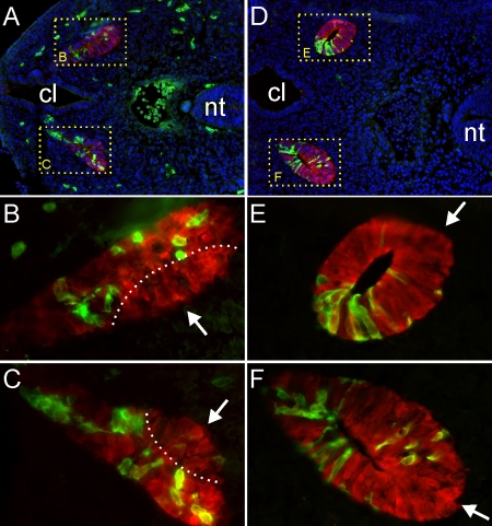
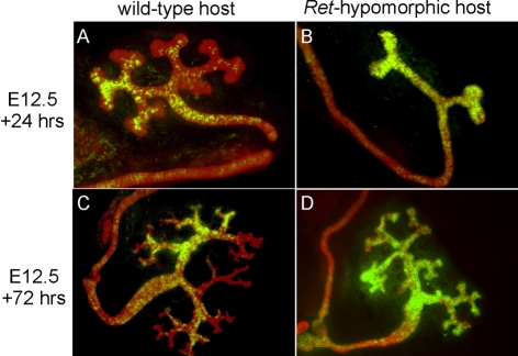
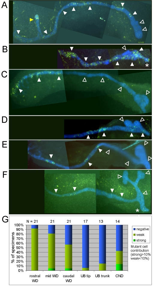
Signaling by the Ret receptor tyrosine kinase promotes cell movements in the Wolffian duct that give rise to the first ureteric bud tip, initiating kidney development. Although the ETS transcription factors Etv4 and Etv5 are known to be required for mouse kidney development and to act downstream of Ret, their specific functions are unclear. Here, we examine their role by analyzing the ability of Etv4 Etv5 compound mutant cells to contribute to chimeric kidneys. Etv4(-/-);Etv5(+/-) cells show a limited distribution in the caudal Wolffian duct and ureteric bud, similar to Ret(-/-) cells, revealing a cell-autonomous role for Etv4 and Etv5 in the cell rearrangements promoted by Ret. By contrast, Etv4(-/-);Etv5(-/-) cells display more severe developmental limitations, suggesting a broad role for Etv4 and Etv5 downstream of multiple signals, which are together important for Wolffian duct and ureteric bud morphogenesis.
Figures

Similar articles
-
Etv4 and Etv5 are required downstream of GDNF and Ret for kidney branching morphogenesis.Nat Genet. 2009 Dec;41(12):1295-302. doi: 10.1038/ng.476. Epub 2009 Nov 8. Nat Genet. 2009. PMID: 19898483 Free PMC article.
-
GDNF/Ret signaling and renal branching morphogenesis: From mesenchymal signals to epithelial cell behaviors.Organogenesis. 2010 Oct-Dec;6(4):252-62. doi: 10.4161/org.6.4.12680. Organogenesis. 2010. PMID: 21220964 Free PMC article.
-
Ret and Etv4 Promote Directed Movements of Progenitor Cells during Renal Branching Morphogenesis.PLoS Biol. 2016 Feb 19;14(2):e1002382. doi: 10.1371/journal.pbio.1002382. eCollection 2016 Feb. PLoS Biol. 2016. PMID: 26894589 Free PMC article.
-
GDNF/Ret signaling and the development of the kidney.Bioessays. 2006 Feb;28(2):117-27. doi: 10.1002/bies.20357. Bioessays. 2006. PMID: 16435290 Review.
-
ETV1, 4 and 5: an oncogenic subfamily of ETS transcription factors.Biochim Biophys Acta. 2012 Aug;1826(1):1-12. doi: 10.1016/j.bbcan.2012.02.002. Epub 2012 Mar 8. Biochim Biophys Acta. 2012. PMID: 22425584 Free PMC article. Review.
Cited by
-
Regulation of Renal Differentiation by Trophic Factors.Front Physiol. 2018 Nov 12;9:1588. doi: 10.3389/fphys.2018.01588. eCollection 2018. Front Physiol. 2018. PMID: 30483151 Free PMC article. Review.
-
FGF-Regulated ETV Transcription Factors Control FGF-SHH Feedback Loop in Lung Branching.Dev Cell. 2015 Nov 9;35(3):322-32. doi: 10.1016/j.devcel.2015.10.006. Dev Cell. 2015. PMID: 26555052 Free PMC article.
-
Etv5 Is Required for Peripheral Nerve Function and the Injury Response.eNeuro. 2025 Jul 23;12(7):ENEURO.0410-20.2025. doi: 10.1523/ENEURO.0410-20.2025. Print 2025 Jul. eNeuro. 2025. PMID: 40701803 Free PMC article.
-
Receptor tyrosine kinases in kidney development.J Signal Transduct. 2011;2011:869281. doi: 10.1155/2011/869281. Epub 2011 Mar 3. J Signal Transduct. 2011. PMID: 21637383 Free PMC article.
-
Nephron Progenitor Maintenance Is Controlled through Fibroblast Growth Factors and Sprouty1 Interaction.J Am Soc Nephrol. 2020 Nov;31(11):2559-2572. doi: 10.1681/ASN.2020040401. Epub 2020 Aug 4. J Am Soc Nephrol. 2020. PMID: 32753399 Free PMC article.
References
-
- Airik R., Kispert A. (2007). Down the tube of obstructive nephropathies: the importance of tissue interactions during ureter development. Kidney Int. 72, 1459-1467 - PubMed
-
- Basson M. A., Akbulut S., Watson-Johnson J., Simon R., Carroll T. J., Shakya R., Gross I., Martin G. R., Lufkin T., McMahon A. P., et al. (2005). Sprouty1 is a critical regulator of GDNF/RET-mediated kidney induction. Dev. Cell 8, 229-239 - PubMed
-
- Basson M. A., Watson-Johnson J., Shakya R., Akbulut S., Hyink D., Costantini F. D., Wilson P. D., Mason I. J., Licht J. D. (2006). Branching morphogenesis of the ureteric epithelium during kidney development is coordinated by the opposing functions of GDNF and Sprouty1. Dev. Biol. 299, 466-477 - PubMed
-
- Batourina E., Choi C., Paragas N., Bello N., Hensle T., Costantini F. D., Schuchardt A., Bacallao R. L., Mendelsohn C. L. (2002). Distal ureter morphogenesis depends on epithelial cell remodeling mediated by vitamin A and Ret. Nat. Genet. 32, 109-115 - PubMed
-
- Batourina E., Tsai S., Lambert S., Sprenkle P., Viana R., Dutta S., Hensle T., Wang F., Niederreither K., McMahon A. P., et al. (2005). Apoptosis induced by vitamin A signaling is crucial for connecting the ureters to the bladder. Nat. Genet. 37, 1082-1089 - PubMed
Publication types
MeSH terms
Substances
Grants and funding
LinkOut - more resources
Full Text Sources
Molecular Biology Databases
