

Dissection of the HIV Vif interaction with human E3 ubiquitin ligase
- PMID: 20463065
- PMCID: PMC2898223
- DOI: 10.1128/JVI.00031-10
Dissection of the HIV Vif interaction with human E3 ubiquitin ligase
Abstract
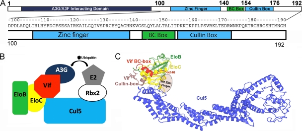
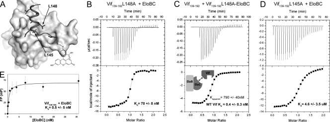
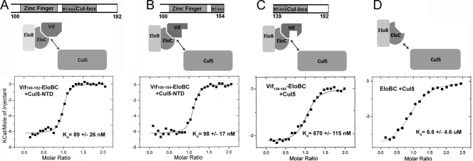
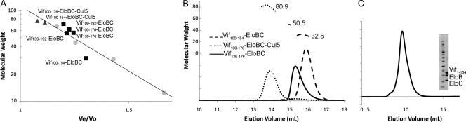
The human immunodeficiency virus type 1 (HIV-1) protein Vif recruits the host E3 ubiquitin ligase, composed of cullin 5 (Cul5), Rbx2, Elongin B, and Elongin C (EloBC), to polyubiquitinate the antiviral protein APOBEC3G. Multiple regions in the C-terminal half of Vif interact with the E3 ligase. We have purified individual regions of Vif and investigated their thermodynamic contributions to the ligase assembly in vitro using isothermal titration calorimetry and fluorescence anisotropy. Our results quantify the high-affinity interactions between the Vif BC box and EloBC and between the Vif zinc finger and Cul5, as well as the modest interaction between the Vif cullin box and Cul5. Our purified Vif constructs also provide direct biochemical evidence that the Vif cullin box, containing the PPLP region, leads to the dimerization of Vif-EloBC complexes but not Cul5-Vif-EloBC complexes.
Figures
Similar articles
-
The SOCS-box of HIV-1 Vif interacts with ElonginBC by induced-folding to recruit its Cul5-containing ubiquitin ligase complex.PLoS Pathog. 2010 Jun 3;6(6):e1000925. doi: 10.1371/journal.ppat.1000925. PLoS Pathog. 2010. PMID: 20532212 Free PMC article.
-
T-cell differentiation factor CBF-β regulates HIV-1 Vif-mediated evasion of host restriction.Nature. 2011 Dec 21;481(7381):376-9. doi: 10.1038/nature10718. Nature. 2011. PMID: 22190036
-
Ubiquitination of APOBEC3G by an HIV-1 Vif-Cullin5-Elongin B-Elongin C complex is essential for Vif function.J Biol Chem. 2005 May 13;280(19):18573-8. doi: 10.1074/jbc.C500082200. Epub 2005 Mar 21. J Biol Chem. 2005. PMID: 15781449
-
The assembly of Vif ubiquitin E3 ligase for APOBEC3 degradation.Arch Pharm Res. 2015 Apr;38(4):435-45. doi: 10.1007/s12272-014-0519-x. Epub 2014 Nov 20. Arch Pharm Res. 2015. PMID: 25408426 Review.
-
Structural insights for HIV-1 therapeutic strategies targeting Vif.Trends Biochem Sci. 2014 Sep;39(9):373-80. doi: 10.1016/j.tibs.2014.07.001. Epub 2014 Aug 12. Trends Biochem Sci. 2014. PMID: 25124760 Free PMC article. Review.
Cited by
-
Protein intrinsic disorder as a flexible armor and a weapon of HIV-1.Cell Mol Life Sci. 2012 Apr;69(8):1211-59. doi: 10.1007/s00018-011-0859-3. Epub 2011 Oct 28. Cell Mol Life Sci. 2012. PMID: 22033837 Free PMC article. Review.
-
Core binding factor beta plays a critical role by facilitating the assembly of the Vif-cullin 5 E3 ubiquitin ligase.J Virol. 2014 Mar;88(6):3309-19. doi: 10.1128/JVI.03824-13. Epub 2014 Jan 3. J Virol. 2014. PMID: 24390320 Free PMC article.
-
Structural perspectives on HIV-1 Vif and APOBEC3 restriction factor interactions.Protein Sci. 2020 Feb;29(2):391-406. doi: 10.1002/pro.3729. Epub 2019 Nov 29. Protein Sci. 2020. PMID: 31518043 Free PMC article. Review.
-
Importance of the proline-rich multimerization domain on the oligomerization and nucleic acid binding properties of HIV-1 Vif.Nucleic Acids Res. 2011 Mar;39(6):2404-15. doi: 10.1093/nar/gkq979. Epub 2010 Nov 13. Nucleic Acids Res. 2011. PMID: 21076154 Free PMC article.
-
Structural analysis of viral infectivity factor of HIV type 1 and its interaction with A3G, EloC and EloB.PLoS One. 2014 Feb 26;9(2):e89116. doi: 10.1371/journal.pone.0089116. eCollection 2014. PLoS One. 2014. PMID: 24586532 Free PMC article.
References
Publication types
MeSH terms
Substances
Grants and funding
LinkOut - more resources
Full Text Sources
Other Literature Sources
