. 2010 Aug;84(15):7730-42.
doi: 10.1128/JVI.02696-09. Epub 2010 May 12.

A spread-deficient cytomegalovirus for assessment of first-target cells in vaccination

Affiliations

A spread-deficient cytomegalovirus for assessment of first-target cells in vaccination

Christian Andreas Mohr et al. J Virol. 2010 Aug.

Abstract

Human cytomegalovirus (HCMV) is a human pathogen that causes severe disease primarily in the immunocompromised or immunologically immature individual. To date, no vaccine is available. We describe use of a spread-deficient murine CMV (MCMV) as a novel approach for betaherpesvirus vaccination. To generate a spread-deficient MCMV, the conserved, essential gene M94 was deleted. Immunization with MCMV-DeltaM94 is apathogenic and protective against wild-type challenge even in highly susceptible IFNalphabetaR(-/-) mice. MCMV-DeltaM94 was able to induce a robust CD4(+) and CD8(+) T-cell response as well as a neutralizing antibody response comparable to that induced by wild-type infection. Endothelial cells were identified as activators of CD8(+) T cells in vivo. Thus, the vaccination with a spread-deficient betaherpesvirus is a safe and protective strategy and allows the linkage between cell tropism and immunogenicity. Furthermore, genomes of MCMV-DeltaM94 were present in lungs 12 months after infection, revealing first-target cells as sites of genome maintenance.


Figures
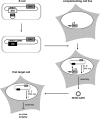
FIG. 1.
FIG. 1.
Concept of inducible trans complementation. In Escherichia coli the BAC pSM3fr-ΔM94 was generated by insertion of the tTA transactivator cassette into pSM3fr, thereby deleting M94. The trans-complementing cell line NT/M94-7 expresses pM94 under the control of the Tet-inducible promoter. Upon transfection with pSM3fr-ΔM94, expression of tTA by the viral genome induces expression of pM94 by the cell, leading to the production of trans-complemented MCMV-ΔM94. This virus is able to infect noncomplementing first-target cells. Due to the lack of the essential gene M94, the release of infectious virus particles is impossible although immediate early (IE), early (E), and late (L) viral gene expression as well as DNA replication (DNA rep) occurs.
FIG. 2.
FIG. 2.
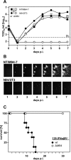
MCMV-ΔM94 is spread deficient, and replication depends on trans complementation of pM94. (A) Parental NIH 3T3 and NT/M94-7 fibroblasts were infected at 0.1 TCID50/cell with MCMV-wt (w) or MCMV-ΔM94M94). At the indicated days, infectious virus in the supernatant was quantified on NT/M94-7 cells by TCID50 endpoint titration. Shown are the means plus SD of titrated duplicates. At day 5 postinfection (p.i.) supernatants were additionally titrated on MEF. No PFU was found in 1 ml supernatant of MCMV-ΔM94-infected NT/M94-7 cells. DL, detection limit. (B) Parental NIH 3T3 and NT/M94-7 fibroblasts were infected with MCMV-Δm157-rec-egfpM94. At the indicated time points EGFP-expressing cells were monitored. hpi, hours p.i. (C) 129.IFNαβR−/− mice (n = 15 for MCMV-ΔM94, n = 8 for MCMV-wt) were infected with 2.5 × 10⁵ TCID50 i.p., and survival was followed for 30 days p.i.
FIG. 3.
FIG. 3.
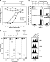
MCMV-ΔM94 induces neutralizing antibodies and T-cell responses. (A) B6 mice were immunized i.p. with 105 TCID50 MCMV-wt (wt) or MCMV-ΔM94M94) or mock infected (PBS). Blood was collected 12 weeks p.i., and virus-neutralizing capacity of the serum was determined using MCMV-luc. Neutralizing antibody (ab) levels of MCMV-ΔM94-immunized mice were significantly lower than antibody levels of MCMV-wt-immunized mice by two-way ANOVA testing (P = 0.04). Values represent the means plus SD of measured serum pools. RLU, relative luciferase units; BG, background. (B) After adoptive transfer of 3 × 10⁵ OT-I CD8+ T cells (top), B6 mice (n = 5) were infected i.p. with 105 TCID50 MCMV-ova (wt-ova) or MCMV-ovaM94M94-ova) or injected with PBS. At days 3, 6, and 8 p.i. flow cytometric analysis was performed on blood for the congenic marker CD45.1 and CD8. After adoptive transfer of 3 × 10⁵ OT-II CD4+ T cells (bottom), B6 mice (n = 5) were infected i.p. as above. At days 3, 6, and 8 p.i. flow cytometric analysis was done on splenocytes for CD90.1 and CD4. Each bar represents the mean plus SD for the indicated group (**, P < 0.01). spec, specific. (C) B6 mice (n = 5) were infected i.p. with 105 TCID50 MCMV-wt (wt), MCMV-ΔM94M94), or UV-irradiated MCMV-wt (wt UV). At day 6 p.i. an in vivo cytotoxicity assay was performed using splenocytes labeled with carboxyfluorescein succinimidyl ester (CFSE) and the indicated viral peptides. Symbols represent the specific lysis activity against the indicated peptide in individual animals. The cross bars indicate the medians of the analyzed groups. The right panel shows an exemplary set of flow cytometric data.
FIG. 4.
FIG. 4.
EC contribute to antiviral CD8+ T-cell stimulation. One day prior to i.p. injection of 105 TCID50 of MCMV-flox-ovaM94M94-flox-ova), MCMV-ovaM94M94-ova), MCMV-wt (wt), or PBS, 3 × 10⁵ congenic OT-I CD8+ T cells were transferred i.v. into B6, Alb-cre, and Tie2-cre mice. At day 6 p.i. a flow cytometric analysis was performed on peripheral blood lymphocytes (PBL) for the congenic marker CD45.1 and CD8. Boxes represent the ratios of OT-I cells per CD8+ cells as a pool of 3 independent experiments and extend from the 25th to the 75th percentile. The lines indicate the medians. Whiskers extend to show the extreme values. The P values were obtained by applying a two-tailed Wilcoxon rank sum test (**, P < 0.01; ***, P < 0.001).
FIG. 5.
FIG. 5.
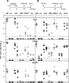
MCMV-ΔM94 protects against challenge with MCMV-wt. B6 mice (n = 5) were immunized (1st) s.c. or i.p. with 105 TCID50 MCMV-wt (wt; solid symbols), MCMV-ΔM94M94; open symbols), Δm01-17+m144-158-MCMV (ΔΔ; dark gray symbols), or PBS (light gray symbols). Virus preparations were UV irradiated before immunization (UV) as indicated. Optionally, mice were boosted (2nd) 2 weeks later with the same dose, route, and virus. Challenge infection was applied i.v. 20 (A) or 4 weeks (B) after priming with 106 PFU MCMV-wt. Five days postchallenge, a plaque assay was performed. Horizontal bars show the medians for the groups. Each symbol represents one individual mouse. DL, detection limit.
FIG. 6.
FIG. 6.
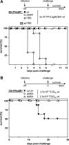
MCMV-ΔM94 protects immunocompromised mice against MCMV-wt challenge. (A) B6.IFNαβR−/− (n = 6) mice were immunized i.p. with 3 × 10⁵ TCID50 MCMV-wt (wt) or MCMV-ΔM94M94). Control groups of B6.IFNαβR−/− (gray circles) or B6 (gray triangles) mice were treated with PBS. Four weeks later challenge infection was performed by i.p. injection of mice with 2 × 10⁵ PFU salivary gland-derived MCMV (sgMCMV-wt), and survival was monitored. (B) 129.IFNαβR−/− mice immunized 4 weeks earlier with 2.5 × 10⁵ TCID50 of MCMV-ΔM94M94; open circles; n = 8), or UV-irradiated MCMV-wt (wt UV; n = 8) were challenged with a lethal dose of MCMV-wt (see Fig. 2C), and survival was monitored. A 10-fold-higher dose of MCMV-wt was applied to mice immunized with MCMV-ΔM94 (n = 7) (open triangles).
FIG. 7.
FIG. 7.
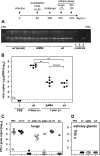
Long-term maintenance of MCMV-ΔM94 genome and protection, B6 mice were infected i.p. with 105 TCID50 MCMV-wt (wt) (n = 5) or MCMV-ΔM94M94) (n = 6). Twelve months p.i. total DNA was extracted from lungs. (A) PCR analysis was performed, obtaining a specific 246-bp fragment of the polymerase gene M54. As controls DNA from lungs 5 days after infection with 105 TCID50 MCMV-wt [wt (acute)] (n = 5), PBS (1), no template (2), or the BAC plasmid pSM3fr (3) was used. MW, molecular weight marker. (B) Quantitative real-time PCR analysis was performed, and viral M54 gene copies per μg genomic DNA were calculated. Each symbol represents one individual mouse. Horizontal bars show the medians of the groups. Genome copy numbers of MCMV-wt (wt) and MCMV-ΔM94M94) are not significantly different (P > 0.05). Both groups are significantly different from mice with acutely infected lungs [wt (acute)] (**, P < 0.01). DL, detection limit. (C and D) B6 mice (n = 5) were immunized i.p. with 105 TCID50 MCMV-wt (wt; solid symbols), MCMV-ΔM94M94; open symbols), Δm01-17+m144-158-MCMV (ΔΔ; dark gray symbols), or PBS (light gray symbols). Virus preparations were UV irradiated before immunization (UV) as indicated. Challenge infection was applied i.v. 1 year after priming with 106 PFU MCMV-wt. The plaque assay was performed 5 days after challenge in lungs (C) and 14 days after challenge in salivary glands (D). Horizontal bars show the medians of the groups. Each symbol represents one individual mouse.

Similar articles

Cited by

References

    1. Adler, S. P., S. E. Starr, S. A. Plotkin, S. H. Hempfling, J. Buis, M. L. Manning, and A. M. Best. 1995. Immunity induced by primary human cytomegalovirus infection protects against secondary infection among women of childbearing age. J. Infect. Dis. 171:26-32. - PubMed
    1. Andrews, D. M., C. E. Andoniou, F. Granucci, P. Ricciardi-Castagnoli, and M. A. Degli-Esposti. 2001. Infection of dendritic cells by murine cytomegalovirus induces functional paralysis. Nat. Immunol. 2:1077-1084. - PubMed
    1. Babu, J. S., J. Thomas, S. Kanangat, L. A. Morrison, D. M. Knipe, and B. T. Rouse. 1996. Viral replication is required for induction of ocular immunopathology by herpes simplex virus. J. Virol. 70:101-107. - PMC - PubMed
    1. Bale, J. F., Jr., S. J. Petheram, I. E. Souza, and J. R. Murph. 1996. Cytomegalovirus reinfection in young children. J. Pediatr. 128:347-352. - PubMed
    1. Balthesen, M., M. Messerle, and M. J. Reddehase. 1993. Lungs are a major organ site of cytomegalovirus latency and recurrence. J. Virol. 67:5360-5366. - PMC - PubMed

Publication types

MeSH terms