. 2010 Jul;84(14):7053-63.
doi: 10.1128/JVI.00737-10. Epub 2010 May 12.

Oligomerization of Ebola virus VP40 is essential for particle morphogenesis and regulation of viral transcription

Affiliations

Oligomerization of Ebola virus VP40 is essential for particle morphogenesis and regulation of viral transcription

T Hoenen et al. J Virol. 2010 Jul.

Abstract

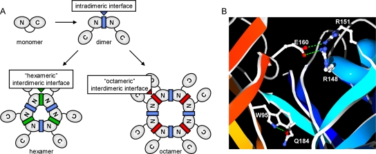
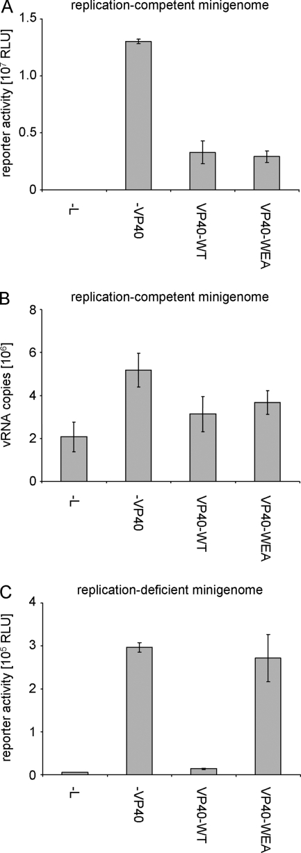
The morphogenesis and budding of virus particles represent an important stage in the life cycle of viruses. For Ebola virus, this process is driven by its major matrix protein, VP40. Like the matrix proteins of many other nonsegmented, negative-strand RNA viruses, VP40 has been demonstrated to oligomerize and to occur in at least two distinct oligomeric states: hexamers and octamers, which are composed of antiparallel dimers. While it has been shown that VP40 oligomers are essential for the viral life cycle, their function is completely unknown. Here we have identified two amino acids essential for oligomerization of VP40, the mutation of which blocked virus-like particle production. Consistent with this observation, oligomerization-deficient VP40 also showed impaired intracellular transport to budding sites and reduced binding to cellular membranes. However, other biological functions, such as the interaction of VP40 with the nucleoprotein, NP, remained undisturbed. Furthermore, both wild-type VP40 and oligomerization-deficient VP40 were found to negatively regulate viral genome replication, a novel function of VP40, which we have recently reported. Interestingly, while wild-type VP40 was also able to negatively regulate viral genome transcription, oligomerization-deficient VP40 was no longer able to fulfill this function, indicating that regulation of viral replication and transcription by VP40 are mechanistically distinct processes. These data indicate that VP40 oligomerization not only is a prerequisite for intracellular transport of VP40 and efficient membrane binding, and as a consequence virion morphogenesis, but also plays a critical role in the regulation of viral transcription by VP40.


Figures

FIG. 1.
FIG. 1.
VP40 oligomeric states. (A) Schematic representation of the different oligomers formed by VP40. Shown are monomers, dimers, hexamers, and octamers, with the N- and C-terminal domains of VP40 indicated. The intradimeric protomer-protomer interface, which is predicted to be similar within hexamers and octamers, is indicated in blue. The interdimeric dimer-dimer interfaces of hexamers and octamers, which are predicted to differ substantially, are indicated in green and red, respectively. (B) Crystal structure of the VP40 intradimeric interface. Polar interactions (shown in green) at the intradimeric interface between two VP40 molecules (depicted in red and blue) in VP40 octamers. Secondary structures are shown in a ribbon representation, and the side chains of interacting amino acids are shown in an all-atom representation. The image was created using SwissPDB-Viewer 3.7 (13) and POV-Ray 3.5.
FIG. 2.
FIG. 2.
W95 and E160 are essential for VP40 homooligomerization. (A) Interaction of VP40 in a mammalian two-hybrid assay. 293 cells were transfected with 500 ng each of plasmids encoding a GAL4 binding domain (pBind) and a VP16 activation domain (pAct) (negative control), pBind-Id and pAct-myo (positive control), or fusion proteins consisting of the GAL4 binding domain or the VP16 domain and VP40-WT or VP40-mutants, as indicated. Five hundred nanograms of a GAL4-driven luciferase reporter construct was also cotransfected. Cells were harvested 48 h after transfection, and luciferase reporter activity, reflecting interaction of VP40, was measured. (B) In vitro oligomerization of VP40. VP40-WT, VP40-WA, VP40-EA, and VP40-WEA were expressed as fusion proteins with glutathione S-transferase (GST) in bacteria and purified via the GST moiety. The GST was subsequently cleaved off using a specific protease, and the purified VP40 was cross-linked using glutaraldehyde. VP40 was detected after SDS-PAGE and Western blotting using VP40-specific antibodies. Monomeric and oligomeric forms of VP40 are labeled. The asterisk indicates uncleaved GST-VP40. (C) Quantitative analysis of cross-linked VP40. The average percentages of the different monomeric and oligomeric forms of VP40 after cross-linking from 3 independent experiments are shown.
FIG. 3.
FIG. 3.
Influence of VP40 oligomerization on production of infectious VLPs. (A) Viral transcription/replication in an iVLP assay. 293 producer cells (p0) were transfected with plasmids encoding all viral structural proteins as indicated, as well as expression plasmids for a minigenome, containing a Renilla luciferase reporter gene, and for T7 polymerase. Seventy-two hours after transfection, reporter activity derived from minigenome replication and transcription in these cells (p0) was determined. Supernatant of these cells was then used for infection of Vero E6 target cells previously transfected with plasmids encoding for NP, VP35, VP30, and L (p1). Seventy-two hours postinfection, reporter activity in these p1 cells was determined. The average percentages and the standard deviation of 3 independent experiments are shown. (B) Production of VLPs. 293 cells were transfected with plasmids encoding VP40 (wild-type [WT] or mutant WA, EA, or WEA, as indicated). Forty-eight hours after transfection, the cell lysate and supernatant were collected. The supernatant was cleared of cellular debris and separated into three fractions. Fraction 1 remained untreated, fraction 2 was treated with proteinase K, and fraction 3 was treated with proteinase K and Triton X-100. After 1 h at 37°C, the proteinase was heat inactivated and samples were subjected to SDS-PAGE, Western blotting, and staining using VP40-specific antibodies. (C) Quantification of released VLPs. The amount of VP40 in VLPs and in the cell lysate was quantified using the Odyssey infrared imaging system (Li-Cor) after Western blotting. Depicted is the average ratio of the signal of proteinase K-resistant VP40 in VLPs (B; VLPs + Proteinase K) divided by the VP40 signal in the cell lysate from three independent experiments. Error bars indicate standard deviation.
FIG. 4.
FIG. 4.
Intracellular localization and membrane association of VP40. (A) Intracellular localization of VP40. HUH-7 cells were transfected with expression plasmids encoding wild-type VP40 (VP40-WT) or homooligomerization-deficient VP40-WEA. Twenty-one hours posttransfection, cells were fixed in 4% paraformaldehyde, permeabilized, and stained using monoclonal mouse anti-VP40 antibodies. As secondary antibodies, rhodamine-coupled anti-mouse antibodies were used, and nuclei were stained using 4′,6-diamidino-2-phenylindole (DAPI). (B) Quantification of VP40 distribution. The phenotypes of cells in 3 independent immunofluorescence experiments as described in panel A were quantified. Shown is the percentage of cells showing predominantly peripheral clusters or predominantly perinuclear clusters. A total of 628 cells were evaluated. The average percentages and the standard deviation of 3 independent experiments are shown. (C) Flotation analysis of VP40. HUH-7 cells were transfected with expression plasmids encoding VP40 (WT or mutant WEA). Thirty-six hours after transfection, cells were lysed and the cell lysate was subjected to flotation analysis using a Nycodenz gradient. After ultracentrifugation, 5 fractions were taken off the gradient, and VP40 in these fractions was visualized by SDS-PAGE and Western blotting using VP40-specific antibodies. Membrane-associated protein is found in the top fraction (fraction 1), whereas soluble protein is located in the bottom fraction (fraction 5). (D) Quantification of flotation analysis. Experiments were performed as described in panel C. Western blot signals were quantified using an Odyssey infrared imaging system. The average and standard deviation of 3 independent experiments are shown.
FIG. 5.
FIG. 5.
Interaction of VP40 and NP. (A) Colocalization of VP40 and NP. myc-tagged NP and VP40 (wild type [WT] or mutant WEA, as indicated) were coexpressed in HUH-7 cells. Twenty-one hours after transfection, cells were fixed in 4% paraformaldehyde, permeabilized, and stained using a monoclonal mouse-anti-VP40 antibody and a polyclonal rabbit-anti-myc antiserum. As secondary antibodies, rhodamine-coupled anti-mouse and fluorescein isothiocyanate-coupled anti-rabbit antibodies were used. Nuclei were stained using 4′,6-diamidino-2-phenylindole (DAPI). (B) Coimmunoprecipitation of NP and VP40. Flag-tagged VP40 (VP40-WT or VP40-WA, -EA, and -WEA mutants, as indicated) and NP were expressed in 293 cells. Forty-eight hours after transfection, cells were lysed and the lysate was subjected to coimmunoprecipitation using anti-Flag agarose. NP and VP40 in both the cell lysate and the immunoprecipitate were detected by Western blotting using a monoclonal mouse-anti-NP antibody and a polyclonal guinea pig-anti-VP40 antiserum. As secondary antibodies, IRDye-680 anti-mouse (shown in green) and IRDye-800 anti-guinea pig (shown in red) antibodies were used. (C) Quantification of coimmunoprecipitation. Experiments were performed as described for panel B. Signals in the cell lysate and the immunoprecipitate were quantified using an Odyssey infrared imaging system and are graphed as the ratio between immunoprecipitate and cell lysate, with VP40-WT set to 1. The average and standard deviation of 3 independent experiments are shown.
FIG. 6.
FIG. 6.
Influence of VP40 homooligomerization on viral minigenome replication and transcription. (A) Role in a classical minigenome assay. 293 cells were transfected with a T7-driven minigenome encoding Renilla luciferase, and expression plasmids for the viral proteins NP, L, VP35, VP30, and VP40 (wild type [VP40-WT] or mutant VP40-WEA, as indicated) and accessory plasmids as described in Materials and Methods. Forty-eight hours after transfection, cells were lysed and reporter activity, reflecting both viral replication and transcription, was measured. The average and standard deviation of 3 independent experiments are shown. RLU, relative light units. (B) Influence of VP40 homooligomerization on minigenome replication. 293 cells were transfected with minigenome assay components as described for panel A. Forty-eight hours after transfection, total RNA from these cells was isolated and subjected to a strand-specific quantitative RT-PCR detecting only negative-sense vRNA copies. The average vRNA copy number and standard deviation from 2 independent experiments are shown. (C) Influence of VP40 homooligomerization on transcription of a replication-deficient minigenome. 293 cells were transfected with minigenome assay components as described for panel A; however, instead of a classical minigenome, a replication-deficient minigenome was used. Forty-eight hours after transfection, cells were lysed and reporter activity, reflecting only viral transcription, was measured. The average and standard deviation of 3 independent experiments are shown.
FIG. 7.
FIG. 7.
Hypothetical model of membrane binding by VP40. (A) Oligomerization-competent VP40. Monomeric VP40 binds to membranes, which leads to a displacement of the C-terminal domain relative to the N-terminal domain and exposure of the oligomerization interface within the N-terminal domain (depicted as a dark gray box). Oligomerization leads to the recruitment of additional membrane-binding domains, stabilizing the interaction of VP40 with the membrane. For simplicity, only two subunits of VP40 oligomers are depicted. (B) Oligomerization-deficient VP40. Oligomerization-deficient VP40 is not able to recruit additional membrane binding domains and quickly dissociates from cellular membranes.

References

    1. Becker, S., C. Rinne, U. Hofsass, H. D. Klenk, and E. Muhlberger. 1998. Interactions of Marburg virus nucleocapsid proteins. Virology 249:406-417. - PubMed
    1. Bjorndal, A. S., L. Szekely, and F. Elgh. 2003. Ebola virus infection inversely correlates with the overall expression levels of promyelocytic leukaemia (PML) protein in cultured cells. BMC Microbiol. 3:6. - PMC - PubMed
    1. Boehmann, Y., S. Enterlein, A. Randolf, and E. Muhlberger. 2005. A reconstituted replication and transcription system for Ebola virus Reston and comparison with Ebola virus Zaire. Virology 332:406-417. - PubMed
    1. Carroll, A. R., and R. R. Wagner. 1979. Role of the membrane (M) protein in endogenous inhibition of in vitro transcription by vesicular stomatitis virus. J. Virol. 29:134-142. - PMC - PubMed
    1. Ciancanelli, M. J., and C. F. Basler. 2006. Mutation of YMYL in the Nipah virus matrix protein abrogates budding and alters subcellular localization. J. Virol. 80:12070-12078. - PMC - PubMed

Publication types

MeSH terms