. 2010 Jul;84(14):7405-11.
doi: 10.1128/JVI.00576-10. Epub 2010 May 12.

Pathogenic hantaviruses Andes virus and Hantaan virus induce adherens junction disassembly by directing vascular endothelial cadherin internalization in human endothelial cells

Affiliations

Pathogenic hantaviruses Andes virus and Hantaan virus induce adherens junction disassembly by directing vascular endothelial cadherin internalization in human endothelial cells

Elena Gorbunova et al. J Virol. 2010 Jul.

Abstract

Hantaviruses infect endothelial cells and cause 2 vascular permeability-based diseases. Pathogenic hantaviruses enhance the permeability of endothelial cells in response to vascular endothelial growth factor (VEGF). However, the mechanism by which hantaviruses hyperpermeabilize endothelial cells has not been defined. The paracellular permeability of endothelial cells is uniquely determined by the homophilic assembly of vascular endothelial cadherin (VE-cadherin) within adherens junctions, which is regulated by VEGF receptor-2 (VEGFR2) responses. Here, we investigated VEGFR2 phosphorylation and the internalization of VE-cadherin within endothelial cells infected by pathogenic Andes virus (ANDV) and Hantaan virus (HTNV) and nonpathogenic Tula virus (TULV) hantaviruses. We found that VEGF addition to ANDV- and HTNV-infected endothelial cells results in the hyperphosphorylation of VEGFR2, while TULV infection failed to increase VEGFR2 phosphorylation. Concomitant with the VEGFR2 hyperphosphorylation, VE-cadherin was internalized to intracellular vesicles within ANDV- or HTNV-, but not TULV-, infected endothelial cells. Addition of angiopoietin-1 (Ang-1) or sphingosine-1-phosphate (S1P) to ANDV- or HTNV-infected cells blocked VE-cadherin internalization in response to VEGF. These findings are consistent with the ability of Ang-1 and S1P to inhibit hantavirus-induced endothelial cell permeability. Our results suggest that pathogenic hantaviruses disrupt fluid barrier properties of endothelial cell adherens junctions by enhancing VEGFR2-VE-cadherin pathway responses which increase paracellular permeability. These results provide a pathway-specific mechanism for the enhanced permeability of hantavirus-infected endothelial cells and suggest that stabilizing VE-cadherin within adherens junctions is a primary target for regulating endothelial cell permeability during pathogenic hantavirus infection.


Figures
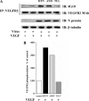
FIG. 1.
FIG. 1.
Pathogenic hantaviruses enhance VEGFR2 phosphorylation responses. HUVECs were mock infected or infected with pathogenic HTNV and ANDV or nonpathogenic TULV at a multiplicity of infection (MOI) of 0.5. Three days postinfection, cells were starved overnight in endothelial basal medium 2 (EBM-2; 0.5% bovine serum albumin [BSA] without growth factors) and subsequently treated with VEGF (100 ng/ml) for 30 min or left untreated (control). Endothelial cells were harvested in lysis buffer (25 mM Tris [pH 7.8], 150 mM NaCl, 2 mM EDTA, 1% NP-40, 10 mM NaF, 2 mM Na3V04, and protease inhibitor cocktail [Sigma]). Cell lysates were clarified by centrifugation and immunoprecipitated (IP) with anti-VEGFR2 rabbit polyclonal antibody (C-1158; Santa Cruz Biotechnology) and protein A/G plus agarose beads (Santa Cruz Biotechnology). Precipitated proteins were fractionated by 7% SDS-PAGE. (A) Immunoblotting (IB) was performed with antibodies against phosphotyrosine (4G10) (Upstate Biotechnology) or anti-VEGFR2 monoclonal antibody (sc-6251; Santa Cruz Biotechnology) and detected using horseradish peroxidase-conjugated anti-mouse and anti-rabbit secondary antibodies (Amersham) and fluorography with enhanced chemiluminescence (Amersham). Control cells were similarly lysed and fractionated by 7% SDS-PAGE, and immunoblotting was performed with antibodies against anti-β-tubulin (Santa Cruz Biotechnology) or antinucleocapsid protein (N protein) as previously described (23). (B) An NIH Image analysis of immunoblotted bands was used to quantitate phosphorylated VEGFR2, and the results are levels normalized to constant amounts of tubulin present on blots.
FIG. 2.
FIG. 2.
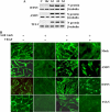
Pathogenic hantaviruses promote VE-cadherin internalization. HUVECs were grown on gelatin-coated coverslips and mock infected or infected with HTNV, ANDV, and TULV at an MOI of 0.5. (A) Three days postinfection, cells were lysed with radioimmunoprecipitation assay buffer (0.1% SDS) and anti-β-tubulin (Santa Cruz Biotechnology), and viral nucleocapsid levels (antinucleocapsid rabbit polyclonal serum [23]) were evaluated by Western blot analysis, as described in the legend to Fig. 1 (46). (B) Three days postinfection, cells were starved overnight in EBM-2 (0.5% BSA without growth factors) and subsequently treated with VEGF (100 ng/ml for 30 min) or mock treated (control). Endothelial cells were subsequently incubated with anti-VE-cadherin antibody, clone BV9 (specific for the extracellular domain of human VE-cadherin; Santa Cruz Biotechnology), in ice-cold EBM-2 (0.5% BSA) at 4°C for 1 h. Unbound antibody was removed by washing coverslips with ice-cold PBS, and cells were shifted to a 37°C CO2 incubator for 1 h to permit VE-cadherin internalization (17, 18). Cells were left untreated or washed with a mild acid solution (2 mM PBS-glycine, pH 2.0; three times for 5 min) to remove VE-cadherin antibody that was not internalized (17, 18). Cells were washed with PBS, fixed in 4% paraformaldehyde (10 min), permeabilized with 0.25% Triton X-100 (5 min), and incubated with secondary anti-mouse FITC-labeled antibody (Jackson Labs) for 1 h (17, 18). Coverslips were mounted using the SlowFade kit (Molecular Probes) and examined using an Olympus IX51 fluorescence microscope. Results are representative of 3 to 5 independent experiments.
FIG. 3.
FIG. 3.
Quantification of VE-cadherin internalization and endothelial cell permeability. (A) Endothelial cells were infected and treated as described in the legend to Fig. 2B. The internalization of endogenous VE-cadherin was quantified using NIH Image to detect VE-cadherin fluorescence (BV9 and FITC, as described for Fig. 2B). The percentage of cells with at least one group of five or more acid-resistant, VE-cadherin-positive vesicle-like structures (internalized VE-cadherin) was quantitated and compared to that of mock, VEGF-untreated, acid-washed controls (17, 18). Findings are from three independent experiments (n = 500 cells) which were analyzed by analysis of variance, and asterisks indicate a significant difference at a P value of <0.001 with respect to unstimulated cells. Results are expressed as the means ± standard errors of the mean (SEM) of three independent experiments. (B) HUVECs were seeded onto vitronectin-coated (10 μg/ml) Costar Transwell inserts (6.5-mm diameter and 3-μm pore size; Corning) at a cell density of 2 × 10⁴ cells/well and grown in EBM-2 (10% of fetal calf serum). Confluent HUVECs were infected in triplicate with pathogenic HTNV or ANDV or nonpathogenic TULV at an MOI of 0.5 or mock infected. Three days postinfection, cells were starved overnight in EBM-2 (0.5% bovine serum albumin without growth factors). FITC-dextran (0.5 mg/ml) was added to the upper chamber in the presence or absence of VEGF (100 ng/ml, 30 min at 37°C) as previously described (22). The level of FITC-dextran in the lower chamber was quantitated using a Perkin-Elmer fluorimeter (490-nm excitation, 530-nm emission).
FIG. 4.
FIG. 4.
Ang-1 and S1P prevent VE-cadherin internalization in hantavirus-infected HUVECS. HUVECs were mock infected or infected with pathogenic HTNV and ANDV or nonpathogenic TULV at an MOI of 0.5. Three days postinfection, cells were starved overnight in EBM-2 (0.5% bovine serum albumin without growth factors) and pretreated with Ang-1 (50 ng/ml) 5 min prior to VEGF addition (100 ng/ml, 30 min) or treated with S1P (1 mM) and VEGF (100 ng/ml, 30 min) (22). Cells were incubated with anti-VE-cadherin antibody and acid washed, fixed, and stained with secondary FITC-labeled antibody as described in the legend to Fig. 2 (17, 18). The percentage of total acid-washed cells exhibiting VE-cadherin fluorescence is presented (±SEM; n = 500; P < 0.001 versus mock-treated, acid-washed controls).

Similar articles

Cited by

References

    1. Aird, W. C. 2004. Endothelium as an organ system. Crit. Care Med. 32:S271-S279. - PubMed
    1. Berger, M. M., C. Hesse, C. Dehnert, H. Siedler, P. Kleinbongard, H. J. Bardenheuer, M. Kelm, P. Bartsch, and W. E. Haefeli. 2005. Hypoxia impairs systemic endothelial function in individuals prone to high-altitude pulmonary edema. Am. J. Respir. Crit. Care Med. 172:763-767. - PubMed
    1. Borges, E., Y. Jan, and E. Ruoslahti. 2000. Platelet-derived growth factor receptor beta and vascular endothelial growth factor receptor 2 bind to the beta 3 integrin through its extracellular domain. J. Biol. Chem. 275:39867-39873. - PubMed
    1. Bustamante, E. A., H. Levy, and S. Q. Simpson. 1997. Pleural fluid characteristics in hantavirus pulmonary syndrome. Chest 112:1133-1136. - PubMed
    1. Byzova, T. V., C. K. Goldman, N. Pampori, K. A. Thomas, A. Bett, S. J. Shattil, and E. F. Plow. 2000. A mechanism for modulation of cellular responses to VEGF: activation of the integrins. Mol. Cell 6:851-860. - PubMed

Publication types

MeSH terms

LinkOut - more resources