. 2010 May 12;30(19):6538-47.
doi: 10.1523/JNEUROSCI.6250-09.2010.

Tissue plasminogen activator alters intracellular sequestration of zinc through interaction with the transporter ZIP4

Affiliations

Tissue plasminogen activator alters intracellular sequestration of zinc through interaction with the transporter ZIP4

Jaime Emmetsberger et al. J Neurosci. .

Abstract

Glutamatergic neurons contain free zinc packaged into neurotransmitter-loaded synaptic vesicles. Upon neuronal activation, the vesicular contents are released into the synaptic space, whereby the zinc modulates activity of postsynaptic neurons though interactions with receptors, transporters and exchangers. However, high extracellular concentrations of zinc trigger seizures and are neurotoxic if substantial amounts of zinc reenter the cells via ion channels and accumulate in the cytoplasm. Tissue plasminogen activator (tPA), a secreted serine protease, is also proepileptic and excitotoxic. However, tPA counters zinc toxicity by promoting zinc import back into the neurons in a sequestered form that is nontoxic. Here, we identify the zinc influx transporter, ZIP4, as the pathway through which tPA mediates the zinc uptake. We show that ZIP4 is upregulated after excitotoxin stimulation of the mouse, male and female, hippocampus. ZIP4 physically interacts with tPA, correlating with an increased intracellular zinc influx and lysosomal sequestration. Changes in prosurvival signals support the idea that this sequestration results in neuroprotection. These experiments identify a mechanism via which neurons use tPA to efficiently neutralize the toxic effects of excessive concentrations of free zinc.


Figures
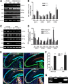
Figure 1.
Figure 1.
ZIP4 increases in expression in the hippocampus during excitotoxic events. A, Representative image of the expression of ZIP isoforms from wt and tPA−/− cortices (Cx) and hippocampi (Hc) was analyzed by RT-PCR. B, Quantification of ZIP isoform expression by densitometry using ImageJ software and comparison using one-way ANOVA. Error bars represent SEM (n = 3 independent experiments). C, Wt and tPA−/− animals were injected unilaterally with kainate (0.75 nmol) or PBS and the mRNA levels of the ZIP isoforms in the cortex and hippocampus (ipsilateral and contralateral to the injection site) were examined. D, Quantification of ZIP isoform expression after kainate injection by densitometry using ImageJ software and comparison using one-way ANOVA followed by Bonferroni's post hoc test. Error bars represent SEM where *p < 0.05, **p < 0.01 (n = 3 independent experiments). E, Wt and tPA−/− animals were unilaterally infused for 24 h with kainate (KA; 0.75 nmol) into the hippocampus, following which the brains were fixed, sectioned, immunostained using anti-ZIP4 (red fluorescence) and anti-NeuN (green, a neuronal marker), and imaged by confocal microscopy. DAPI (blue) denotes nuclei. The CA3 region is marked. Boxed regions are shown at higher magnification in the panels in the inset. DG, Dentate gyrus. F, Comparison of fluorescent intensity of ZIP/NeuN positive cells in the CA3 regions of the ipsilateral and contralateral sides. Quantification was performed by analyzing four 40× images of the CA3 region and measuring the fluorescent intensity using AxioVision 4.6 imaging software. Error bars represent SEM, where **p < 0.0098 by one-tailed Student's t test (n = 3 independent experiments). Scale bar, 20 μm. G, Expression of ZIP1 and ZIP4 in cultured primary hippocampal neurons using RT-PCR. β-Actin was used as control. White arrowhead indicates cells on the ipsilateral side that are positive for nuclear DAPI and NeuN staining and cytoplasmic ZIP4 staining, which presents as yellow or white nuclei surrounded by a red halo.
Figure 2.
Figure 2.
tPA forms a complex with ZIP4 on the cell surface. A, Lysates from HA-mZIP4-expressing HEK293 cells preincubated with or without 10 μg/ml tPA on ice were immunoprecipitated using anti-tPA antibody and immunoblotted using anti-HA antibody to detect HA-tagged ZIP4. B, Lysates of control HEK293 cells and HA-mZIP4-expressing HEK293 cells were preincubated with varied concentrations of tPA, immunoprecipitated with anti-HA, and assayed for tPA activity using zymography. − denotes no addition of tPA. C, Control (injected with PBS) and kainate-injected brains were separated in the injected (ipsi) and uninjected (contra) sides and lysed. The lysates were immunoprecipitated with anti-mZIP4, and assayed for tPA activity using zymography.
Figure 3.
Figure 3.
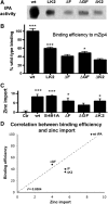
tPA protein domains important for ZIP4 physical and functional interaction. A, HA-mZIP4-expressing HEK293 cells were incubated on ice with 10 μg/ml wt tPA or varied tPA domain mutants, washed, lysed, immunoprecipitated with anti-HA, and assessed for tPA activity via zymography. LK2, Mutant encoding only light catalytic chain and second kringle domain; ΔF, mutant lacking finger domain; ΔGF, mutant lacking growth factor domain; ΔK2, mutant lacking second kringle domain. B, tPA zymographic activity was quantified by densitometry using ImageJ software. The inactive mutant S481A tPA was used as a negative control (data not shown) and its densitometric value (since it is catalytically inactive) was subtracted from the values of all the other recombinant proteins, following which the binding for each mutant was normalized to that observed for the wild-type protein. Error bars represent SEM where *p < 0.05, ***p < 0.001by ANOVA followed by Bonferroni's post hoc test (n = 3 independent experiments). C, HEK293 cells expressing mZIP4 were preloaded with FluoZin-3 and incubated with wt tPA, tPA domain mutants, or catalytically inactive (S481A). Ctr, Control mZIP4-HEK293 cells incubated with media that did not contain recombinant tPA. Zinc import was quantified by fluorometric analysis (ratio A480/A510). D, Correlation between zinc import and ZIP4 binding efficiency by different tPA recombinant proteins.
Figure 4.
Figure 4.
Zinc uptake is augmented in the presence of tPA. HEK cells and mZIP4-expressing HEK cells preloaded with FluoZin-3 were incubated with zinc in the presence or absence of 10 μg/ml tPA. The amount of zinc uptake was quantified by fluorometry (ratio A480/A510).
Figure 5.
Figure 5.
tPA enhances zinc uptake in hippocampal neurons. A, Hippocampal neurons loaded with FluoZin-3 were incubated 0, 10, 35, and 50 μm zinc and/or tPA (10 μg/ml). Quantification of intracellular zinc was performed by fluorometric analysis (A480/A510) at 2 h, and the values are expressed as arbitrary units. The membrane-permeable zinc chelator TPEN was then added and the cells reimaged. The import of zinc is presented in arbitrary units. B, Two-photon imaging of wt hippocampal neurons (naive, treated with tPA, with 50 μm zinc, or with both zinc and tPA) loaded with FluoZin-3 (green) and counterstained against cyt C (red). Arrows indicate coincident fluorescence of FluoZin3 and cyt C. Error bars represent SEM, where **p < 0.01, ***p < 0.001 by two-way ANOVA followed by Bonferroni's post hoc test (4 neurons of each experimental condition were quantified from n = 3 independent experiments). C, Organotypic hippocampal slices from wild-type and tPA−/− mice treated with 50 μm zinc and loaded with Fluozin-3 (green) to assess zinc uptake. Scale bar, 20 μm (n = 3 independent experiments, 3 slices/experiment). The Fluozin-3 fluorescence intensity of the whole slice was quantified, divided by the slice surface area and was analyzed using AxioVision 4.6 image software. Wt slices exhibited 0.58 arbitrary units of fluorescence/surface area compared with 0.35 units for the tPA−/− slices (p = 0.2232 by one-tailed Student's t test).
Figure 6.
Figure 6.
tPA increases vesicular uptake of ZIP4 into lysosomes in ZIP4-expressing HEK293 cells. Confocal images of mZIP4-expressing HEK293 cells incubated with increasing concentrations of zinc in the presence or absence of tPA. A, The presence of mZIP4 was visualized with an anti-HA antibody (red); actin was visualized using phalloidin (green), and nuclei were marked with DAPI (blue). B, Fluorescent intensity of intracellular ZIP4 was quantified using the LSM imaging software. The experiments were repeated at least five times and 30 cells quantified for each condition. Error bars represent SEM, where *p < 0.05, ***p < 0.001 by ANOVA followed by Bonferroni's post hoc test. Scale bar, 20 μm. C, mZIP4 cells were incubated with 50 μm zinc or zinc and tPA. The cells were stained for ZIP4 (red) using the anti-HA antibody, and the lysosomal marker LAMP-1 (green). n = 3 individual experiments.
Figure 7.
Figure 7.
Proapoptotic signals are altered in the presence of tPA. A, Quantitative immunoblot analyses of proteins involved in cell death due to zinc toxicity were performed on HEK cell lysates. mZIP4 HEK cells were treated with various concentrations of zinc with or without tPA and the expression of ERK, p-ERK, JNK, p-JNK, p38, p-p38, AKT, and p-AKT assessed. B, Quantitative measurements of kinase phosphorylation were performed using the Odyssey 2.1 software and the data were plotted as a ratio of the pixel volume of the phosphorylated kinase over the pixel volume of total kinase. For statistical analysis, zinc treatment was compared with zinc plus tPA treatment. Error bars represent SEM, where *p < 0.05, **p < 0.01, ***p < 0.001 by ANOVA followed by Bonferroni's post hoc test (n = 3 independent experiments).

Similar articles

Cited by

References

    1. Andrade-Gordon P, Strickland S. Interaction of heparin with plasminogen activators and plasminogen: effects on the activation of plasminogen. Biochemistry. 1986;25:4033–4040. - PubMed
    1. Belloni-Olivi L, Marshall C, Laal B, Andrews GK, Bressler J. Localization of zip1 and zip4 mRNA in the adult rat brain. J Neurosci Res. 2009;87:3221–3230. - PMC - PubMed
    1. Choi DW, Koh JY. Zinc and brain injury. Annu Rev Neurosci. 1998;21:347–375. - PubMed
    1. Cornford EM, Nguyen EV, Landaw EM. Acute upregulation of blood-brain barrier glucose transporter activity in seizures. Am J Physiol Heart Circ Physiol. 2000;279:H1346–H1354. - PubMed
    1. Coulter D. Mossy fiber zinc and temporal lobe epilepsy: pathological association with altered “epileptic” gamma-aminobutyric acid A receptors in dentate granule cells. Epilepsia. 2000;41(Suppl 6):S96–S99. - PubMed

Publication types

MeSH terms

LinkOut - more resources