. 2010 May 12;30(19):6595-606.
doi: 10.1523/JNEUROSCI.4932-09.2010.

Guidance of postural motoneurons requires MAPK/ERK signaling downstream of fibroblast growth factor receptor 1

Affiliations

Guidance of postural motoneurons requires MAPK/ERK signaling downstream of fibroblast growth factor receptor 1

Prabakaran Soundararajan et al. J Neurosci. .

Abstract

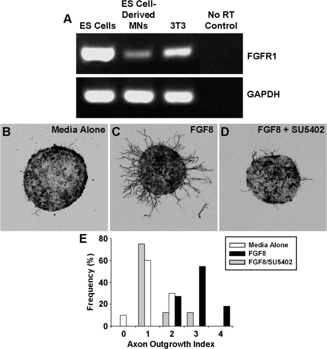
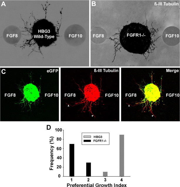
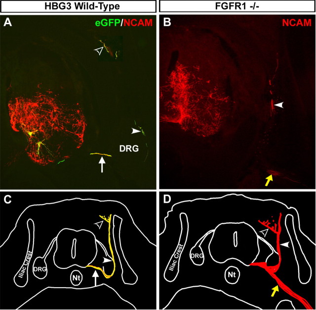
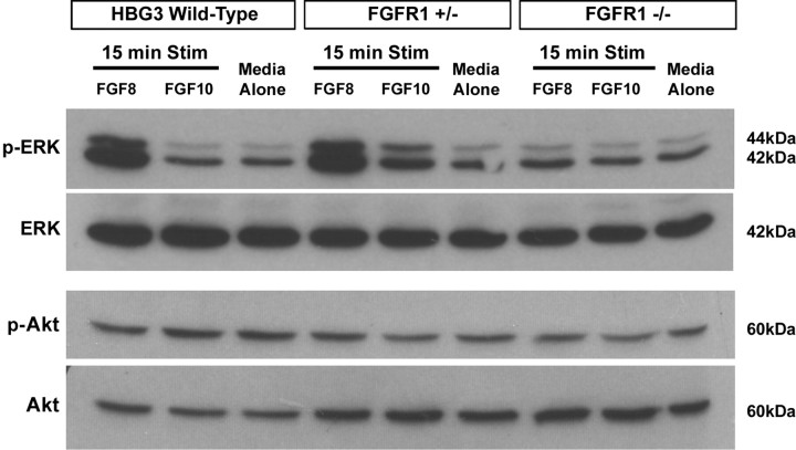
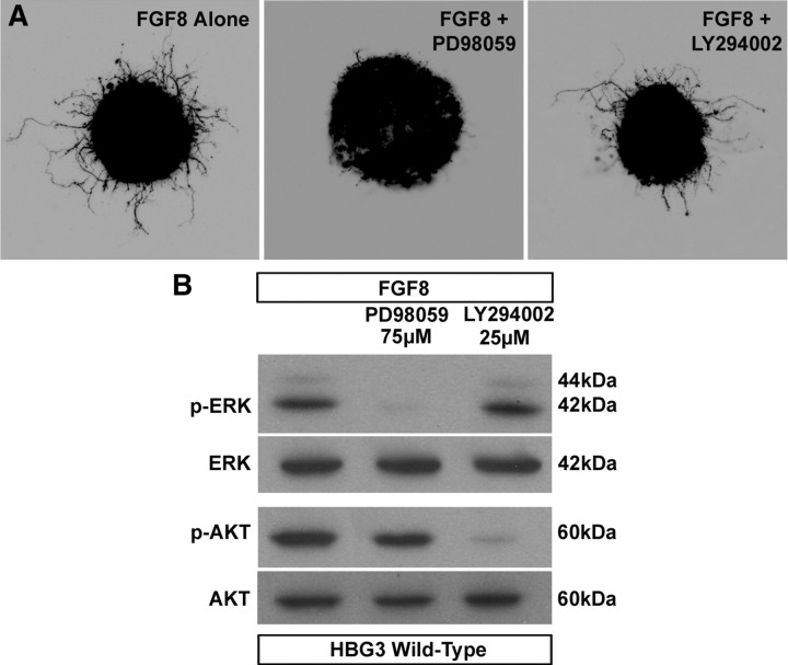
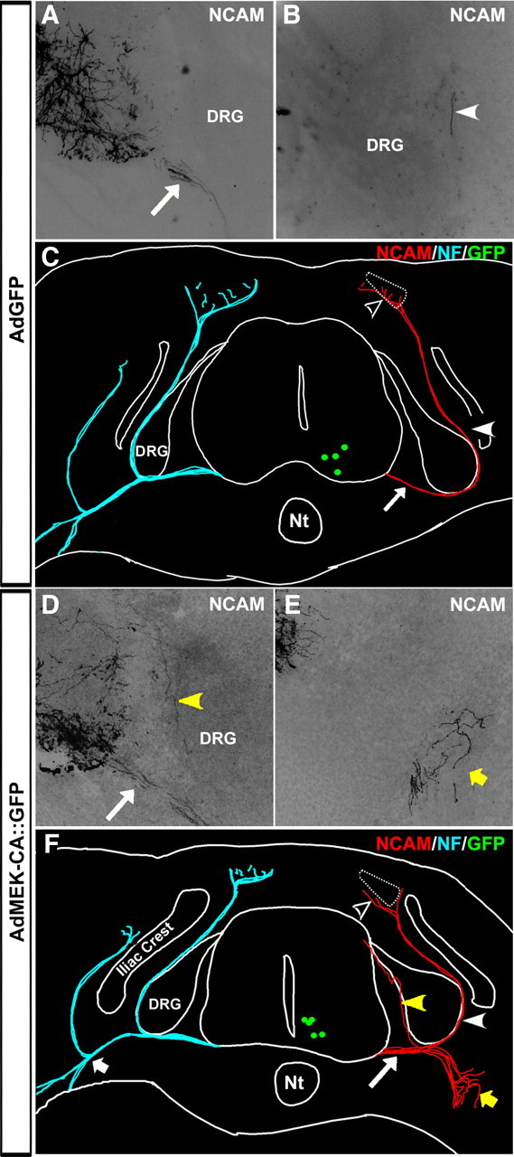
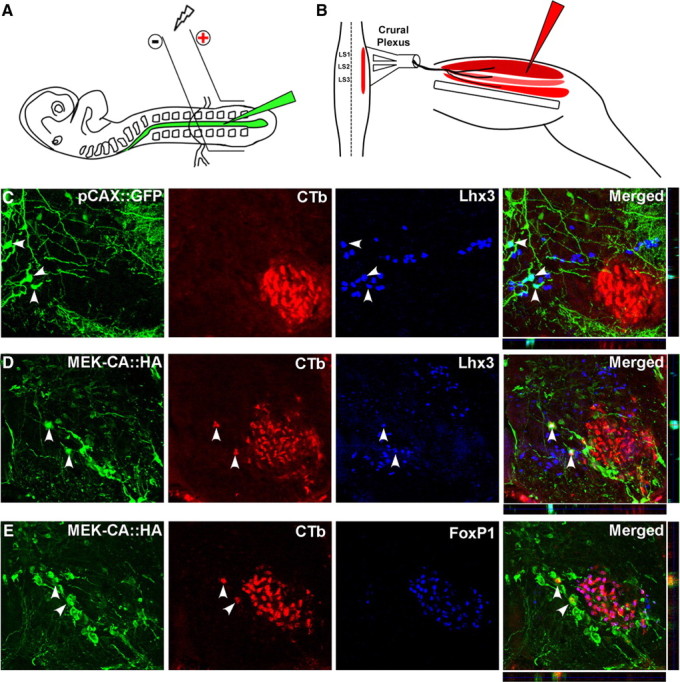
Identification of intracellular signaling pathways necessary for appropriate axon guidance is challenging because many CNS populations used to study these events contain multiple cell types. Here, we resolve this issue by using mouse embryonic stem (ES) cells that were directed to differentiate into a population of motoneurons that exclusively innervate epaxial muscles [medial median motor column (MMCm) motoneurons]. These ES cell-derived MMCm motoneurons, like their endogenous counterparts, express fibroblast growth factor receptor 1 (FGFR1) and selectively extend axons toward the epaxial trophin FGF8. Unlike wild-type MMCm motoneurons, FGFR1(-/-) MMCm motoneurons show guidance defects when transplanted into the neural tube of chick embryos. Furthermore, activation of FGFR1 selectively signals through mitogen-activated protein kinase/extracellular signal-regulated kinase (MAPK/ERK) for appropriate guidance in vitro, whereas overexpression of constitutively active MAPK/ERK in transplanted, or endogenous chick, MMCm cells causes guidance defects in vivo. These results indicate that MAPK/ERK activation downstream of FGFR1 is necessary for MMCm motor axon guidance and that ES cell-derived neurons provide an important tool for dissecting intracellular pathways required for axon guidance.


Figures

Figure 1.
Figure 1.
ES cell-derived MMCm motoneurons express FGFR1 and readily extend axons when exposed to the trophin FGF8. A, RT-PCR analysis revealed that ES cells, ES cell-derived MMCm motoneurons, and 3T3 cells express mRNA for FGFR1. Negative control included RNA samples not subjected to reverse transcription (no RT). GAPDH, Glyceraldehyde-3-phosphate dehydrogenase. B–D, ES cell-derived MMCm motoneurons extended fewer neurites when cultured for 24 h in 3-D collagen gels (n = 11 for each condition) containing media alone (B) compared with those cultured with FGF8 (100 ng/ml) (C). FGF8 (100 ng/ml) induced neurite outgrowth was dramatically attenuated when SU5402 (12.5 μm), an FGFR1/FGFR3 antagonist, was added to the cultures (D). Pictures are z-stack collapsed confocal images in which the green fluorescence is shown as inverted black and white. E, Quantification of neurite outgrowth, using an outgrowth index [for details, see Materials and Methods and supplemental Fig. 1 (available at www.jneurosci.org as supplemental material)]. ANOVA of the quantified growth scores revealed that FGF8-treated ES cell-derived motoneurons had significantly more outgrowth compared with media alone (p < 0.05) or cells treated with FGF8 and SU5402 (p < 0.05). Outgrowth from the latter two conditions was not significantly different from one another.
Figure 2.
Figure 2.
ES cell-derived MMCm motoneurons preferentially extend neurites toward FGF8 in 3-D collagen gels. A, B, ES cell-derived MMCm motoneurons preferentially extended eGFP+ neurites toward FGF8-loaded beads after 24 h in culture, although they were equidistant to FGF10-loaded beads (A). ES cell-derived motoneurons lacking FGFR1−/−, stained for β-III tubulin, did not show preference for either FGF8 or FGF10. Both images are a z-stack collapsed confocal image shown as an inverted black and white of the fluorescence. C, z-stack collapsed confocal images of the eGFP+ ES cell-derived motoneurons, shown in A, immunolabeled for β-III tubulin. Merged image shows that the vast majority of neurites extending into the collagen are eGFP+ and β-III tubulin+. Two eGFP/β-III tubulin+ neurites are indicated with arrowheads. D, Quantification of neurite outgrowth, using a preferential neurite outgrowth index (supplemental Fig. 2, available at www.jneurosci.org as supplemental material). Wild-type ES cell-derived motoneurons extended more neurites toward FGF8 compared with motoneurons lacking FGFR1 (ANOVA, p < 0.05; n = 6 for each condition).
Figure 3.
Figure 3.
Transplanted ES cell-derived MMCm motoneurons lacking FGFR1 exhibit axon guidance defects. A, Cross section through the neural tube of an HH stage 31 (E7) chick embryo transplanted with HBG3 eGFP+ ES cell-derived motoneurons 5 d previously. Section was immunolabeled with a mouse-specific NCAM antibody (red) to visualize transplanted motoneurons and interneurons. Neurons coexpressing eGFP and mouse NCAM appear yellow. Transplanted motoneurons migrated to the MMCm (2 yellow cells), extended axons out of the spinal cord through the ventral root (arrow), and then projected around the DRG as part of the dorsal ramus (arrowhead) before innervating the epaxial muscle (open arrowhead). The NCAM+/eGFP neurons (red axons) are presumably transplanted interneurons that differentiated when the ES cells were treated with RA/Shh. As expected, these neurons branched extensively within the ventral neural tube, but none of them extended axons out of the spinal cord. B, Cross section through the neural tube of an HH stage 31 chick embryo transplanted 5 d previously with ES cell-derived MMCm motoneurons lacking FGFR1−/−. The section was immunostained for mouse-specific NCAM to visualize transplanted cells. Although some FGFR1−/− motor axons correctly navigated to the epaxial muscle as part of the dorsal ramus (arrowhead), many axons incorrectly projected toward the limb (yellow arrow). C, Neurolucida reconstruction shows that all of the transplanted motoneurons in the chick embryo shown in A projected axons out of the spinal cord through the ventral root (arrow) and then dorsally as part of the dorsal ramus (arrowhead). Epaxial muscle is indicated with dotted lines. D, Neurolucida reconstruction of the embryo in B shows that some transplanted ES cell-derived MMCm FGFR1−/− motoneurons projected to the epaxial muscle as part of the dorsal ramus (arrowhead), whereas others incorrectly extended toward the limb (yellow arrow). Nt, Notochord.
Figure 4.
Figure 4.
FGF8 phosphorylates ERK in motoneurons derived from HBG3 and FGFR1+/− but not FGFR1−/− ES cells. A representative Western blot loaded with equal amounts of whole-cell lysate from motoneurons derived from HBG3, FGFR1+/−, and FGFR1−/− ES cells treated for 15 min with FGF8 (50 ng/ml), FGF10 (50 ng/ml), or media alone. p-ERK (1st row) and p-Akt (3rd row). Blots were reprobed with an antibody against total ERK (2nd row) or total Akt (4th row).
Figure 5.
Figure 5.
Inhibition of the MAPK/ERK signaling pathway attenuates FGF8-mediated axonal outgrowth. A, z-stack collapsed confocal images of HBG3 ES cell-derived MMCm motoneurons treated for 12 h with FGF8 alone or in the presence of 75 μm PD98059 or 25 μm LY294002. Axonal outgrowth was dramatically attenuated in cultures containing FGF8 and PD98059 compared with those treated with FGF8 alone or with FGF8 and LY294002. Pictures are shown as inverted black and white images of the green fluorescence. B, Western blot analysis shows that the concentrations of PD98058 and LY294002 used in the outgrowth assays inhibited phosphorylation of ERK and Akt, respectively. Neither inhibitor blocked phosphorylation of the kinase in the other pathway.
Figure 6.
Figure 6.
Constitutively activating MEK in transplanted ES cell-derived MMCm motoneurons causes axonal misprojections. A, B, Cross sections of the ventral horn (A) and DRG (B) from an HH stage 31 chick embryo transplanted with wild-type 129 ES cell-derived MMCm motoneurons infected with AdGFP. Sections were immunostained with an antibody specific to mouse NCAM. Transplanted AdGFP-infected MMCm motor axons extended out of the spinal cord through the ventral root (A, arrow) and around the DRG, where they formed part of the dorsal ramus (B, arrowhead). C, Neurolucida reconstruction of the embryo in A and B shows that all of the transplanted AdGFP-infected motor axons grew out of the neural tube through the ventral root (arrow), formed part of the dorsal ramus (arrowhead), and projected into the epaxial muscle (open arrowhead). For comparison, the projection patterns of the endogenous neurofilament (NF)-positive chick neurons are shown in blue on the contralateral side. Green dots represent transplanted motoneurons still expressing GFP at this stage in development. D, E, Cross sections of the ventral horn (D) and ventral nerve (E) from an HH stage 31 chick embryo transplanted with ES cell-derived MMCm motoneurons infected with AdMEK-CA::GFP. Sections stained as in A and B. Axons from transplanted AdMEK-CA-infected MMCm motoneurons correctly extended out of the spinal cord through the ventral root (D, arrow). However, some axons misprojected into the DRG (D, yellow arrowhead), whereas others incorrectly extended toward the limb, where they defasciculated and wandered at the point where the nerve trifurcates into three branches (E, yellow arrow). F, Neurolucida reconstruction of the embryo in D and E shows AdMEK-CA::GFP-infected motor axons extending out of the neural tube through the ventral root (arrow). Some axons entered the DRG (yellow arrowhead) or misprojected toward the limb, where the branched extensively (yellow arrow). A few AdMek-CA::GFP-infected motoneurons correctly extended axons dorsally (arrowhead) to the epaxial muscle (open arrowhead). Projection patterns of the endogenous neurofilament-positive chick neurons are shown in blue on the contralateral side. Short arrow indicates site of ventral nerve trifurcation. Green dots represent transplanted motoneurons. Nt, Notochord.
Figure 7.
Figure 7.
Constitutively activating MEK in MMCm motoneurons leads to axonal misprojections. A, B, Schematic representation showing the location of electrodes used in electroporations (A) and muscle group injected with Alexa Fluor 594-conjugated CTb (B). C, Immunolabeling and confocal imaging revealed that none of the pCAX::GFP-transfected neurons (green panel; arrowheads) expressing Lhx3 (blue panel; arrowhead) contained CTb (Alexa Fluor 594-conjugated cholera toxin B used to back-label neurons) (red panel). Individual sections in both the x–z (horizontal strip, below merged image) and y–z (vertical strip, beside merged panel) orthogonal planes confirmed colocalization of GFP and Lhx3 (arrowheads in merged panel). D, Immunolabeling and confocal imaging showed several HA-immunopositive (green panel) and Lhx3-immunopositive (blue panel) cells containing CTb (red panel; arrowheads). Imaging in both the x–z and y–z orthogonal planes confirmed colocalization of the three stains. E, Confocal imaging showed that none of the HA+ cells (green panel; arrowhead) containing CTb (red panel; arrowhead) coexpressed the LMC marker FoxP1 (blue panel and merged).

Similar articles

Cited by

References

    1. Atwal JK, Massie B, Miller FD, Kaplan DR. The TrkB-Shc site signals neuronal survival and local axon growth via MEK and P13-kinase. Neuron. 2000;27:265–277. - PubMed
    1. Barres BA, Silverstein BE, Corey DP, Chun LL. Immunological, morphological, and electrophysiological variation among retinal ganglion cells purified by panning. Neuron. 1988;1:791–803. - PubMed
    1. Campbell DS, Holt CE. Apoptotic pathway and MAPKs differentially regulate chemotropic responses of retinal growth cones. Neuron. 2003;37:939–952. - PubMed
    1. Colamarino SA, Tessier-Lavigne M. The axonal chemoattractant netrin-1 is also a chemorepellent for trochlear motor axons. Cell. 1995;81:621–629. - PubMed
    1. Colvin JS, Bohne BA, Harding GW, McEwen DG, Ornitz DM. Skeletal overgrowth and deafness in mice lacking fibroblast growth factor receptor 3. Nat Genet. 1996;12:390–397. - PubMed

Publication types

MeSH terms

Substances