. 2010 May 12;30(19):6620-34.
doi: 10.1523/JNEUROSCI.5778-09.2010.

Identification of Erbin interlinking MuSK and ErbB2 and its impact on acetylcholine receptor aggregation at the neuromuscular junction

Affiliations

Identification of Erbin interlinking MuSK and ErbB2 and its impact on acetylcholine receptor aggregation at the neuromuscular junction

Luca Simeone et al. J Neurosci. .

Abstract

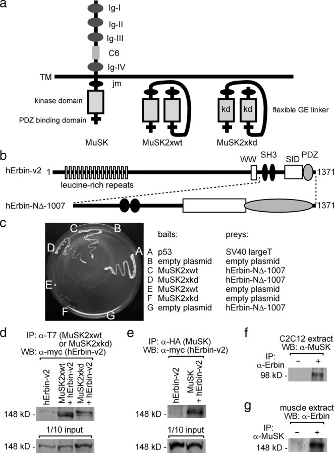
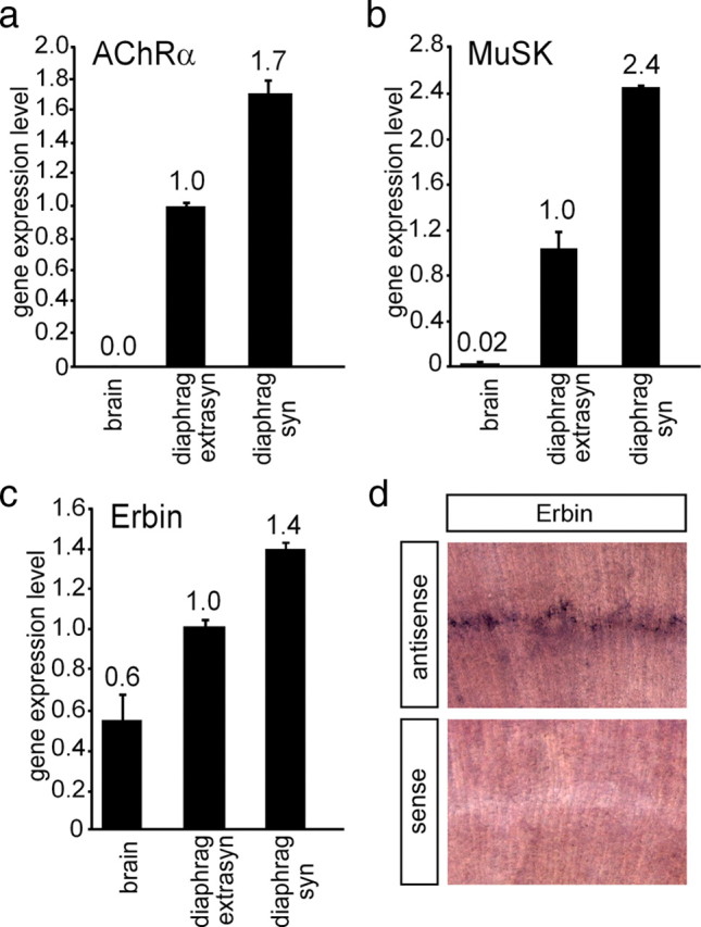
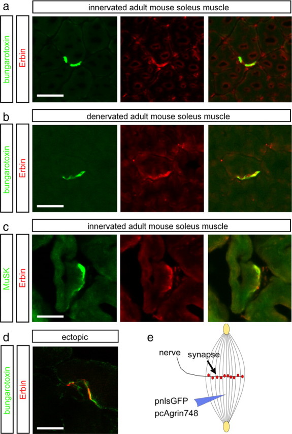
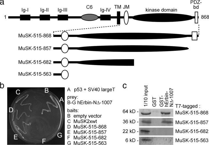
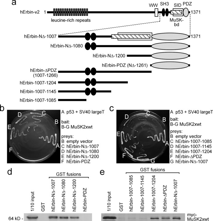
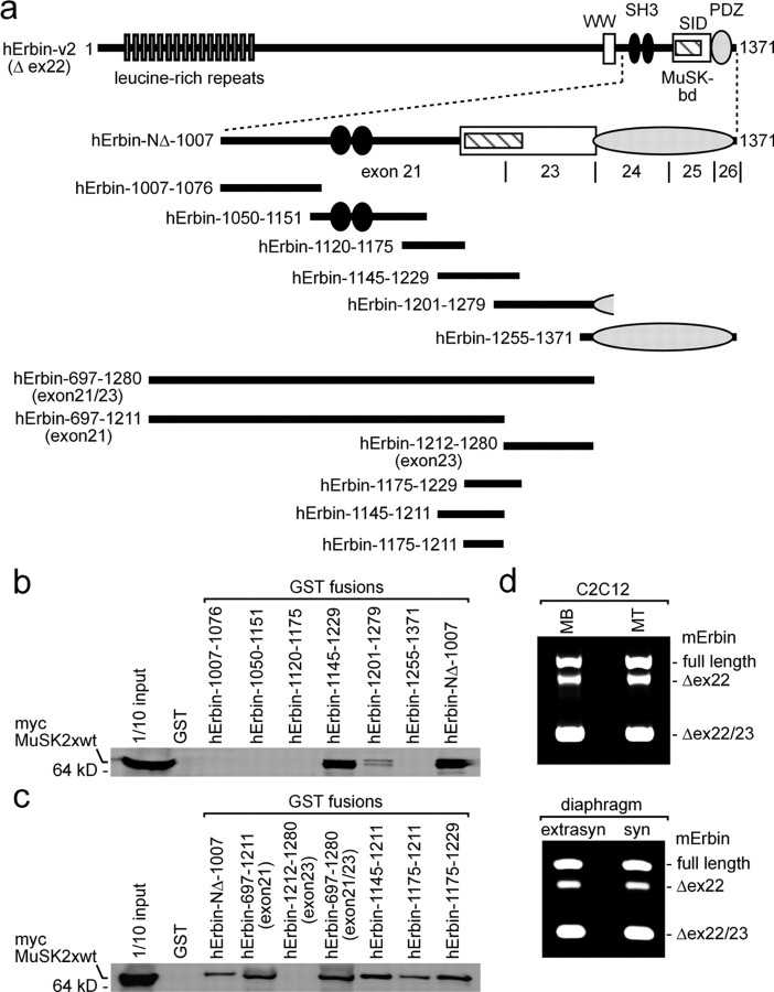
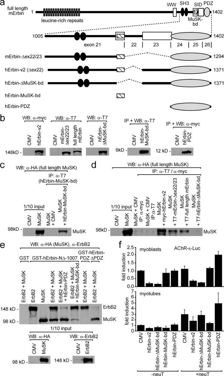
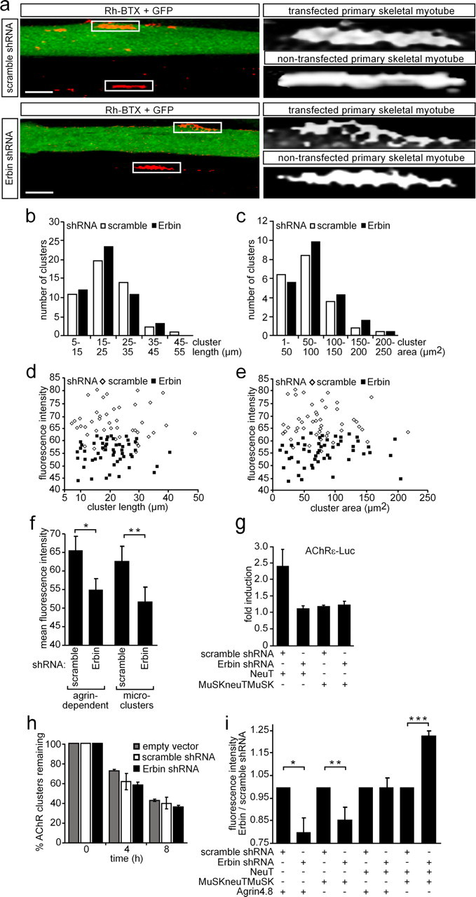
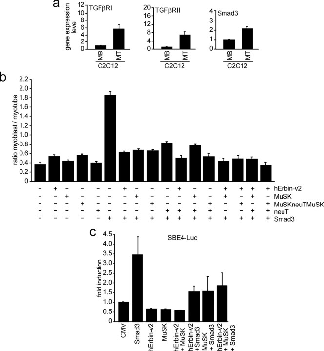
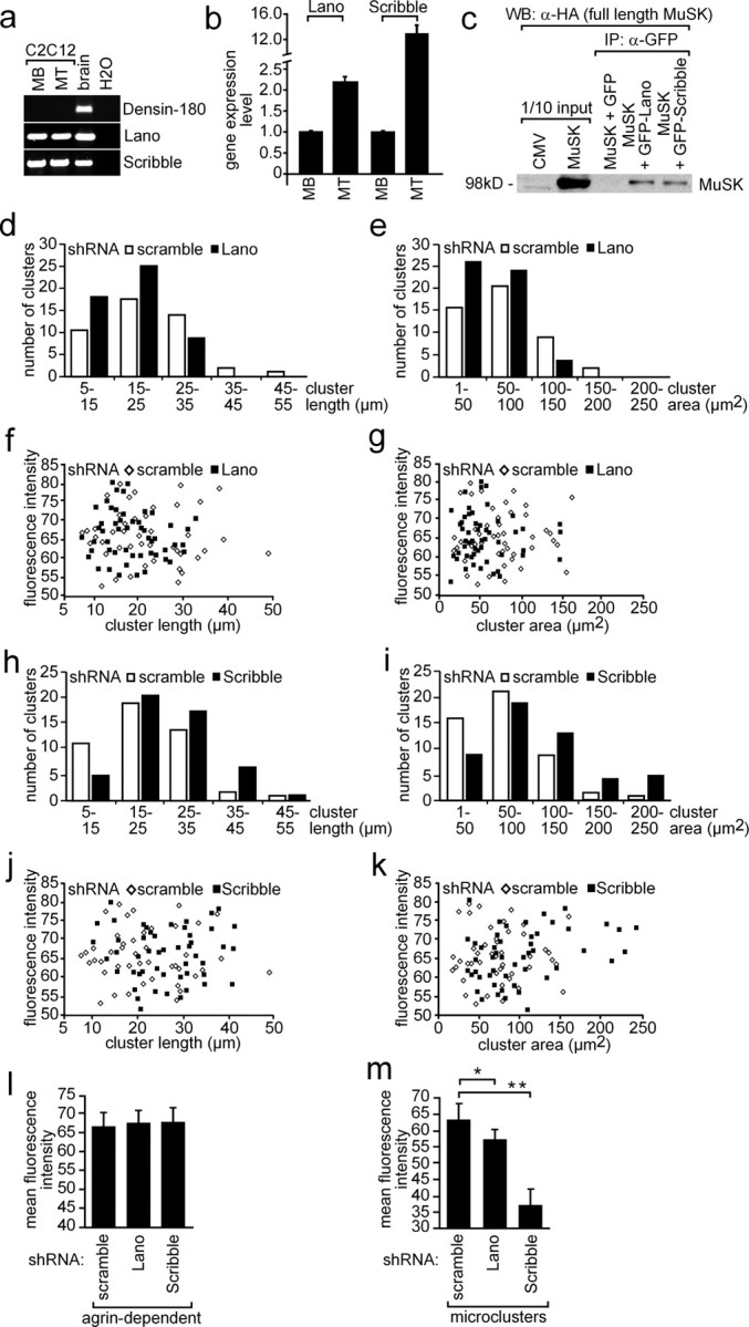
Erbin, a binding partner of ErbB2, was identified as the first member of the LAP family of proteins. Erbin was shown at postsynaptic membranes of the neuromuscular junction (NMJ) or in cultured C2C12 myotubes (1) to be concentrated, (2) to regulate the Ras-Raf-Mek pathway, and (3) to inhibit TGF-beta signaling. In the CNS, Erbin interacts with PSD-95. Furthermore, agrin-MuSK signaling initiates formation of AChR aggregates at the postsynaptic membrane. In search of proteins interacting with MuSK, we identified Erbin as a MuSK binding protein. We verified the interaction of MuSK with Erbin, or both concomitantly with ErbB2 by coimmunoprecipitation, and we mapped the interacting epitopes between Erbin and MuSK. We demonstrated elevated mRNA levels of Erbin at synaptic nuclei and colocalized Erbin and MuSK at postsynaptic membranes. We identified several Erbin isoforms at the NMJ, all of which contained the MuSK binding domain. By knocking down Erbin, we observed agrin-dependent AChR aggregates on murine primary skeletal myotubes and C2C12 cells, and in the absence of agrin, microclusters, both of significantly lower density. Complementary, AChR-epsilon-reporter expression was reduced in myotubes overexpressing Erbin. We show that myotubes also express other LAP protein family members, namely Scribble and Lano, and that both affect physical dimensions of agrin-dependent AChR aggregates and density of microclusters formed in the absence of agrin. Moreover, MuSK-Erbin-ErbB2 signaling influences TGF-beta signaling. Our data define the requirement of Erbin on the cross talk between agrin and neuregulin signaling pathways at the NMJ.


Figures

Figure 1.
Figure 1.
Erbin interacts with MuSK. a, Scheme of wild-type MuSK and two MuSK mutants (MuSK2xwt, MuSK2xkd). The mutants are generated by linking two intracellular parts of MuSK together via a peptide linker, thereby imitating the dimeric active intracellular part of wild-type MuSK. Each of the mutants was used for yeast two-hybrid. The term kd reflects the use of a kinase-defective MuSK mutant. Ig, Ig-like domain; C6, cysteine-rich region; TM, transmembrane segment; JM, juxtamembrane region. b, Illustration of the epitopes of the human Erbin-variant2 (hErbin-v2) (Favre et al., 2001). Below the scheme of human Erbin-v2, the area of Erbin interacting with MuSK is drawn (hErbin-NΔ-1007). SID, Smad-interacting domain; WW, Trp-Trp; SH3, Src homology domain 3. c, The growth of different yeast clones on agar lacking histidine and adenine is shown. Note that yeast cells are able to grow if they contain either MuSK2xwt or MuSK2xkd as bait plasmids and the C-terminal part of human Erbin (hErbin-NΔ-1007) as prey plasmid, or if they contain p53 and SV40 large T antigen (positive control). d, e, Extracts of transiently transfected HEK293 cells were used to precipitate MuSK2xwt, MuSK2xkd, or full-length hemagglutinin (HA)-tagged MuSK. As a consequence human Erbin-v2 was coprecipitated. The amount corresponding to 1/10 of the input was ascertained by Western blot. f, g, Precipitation of endogenous Erbin or MuSK proteins from C2C12 cells or hindlimb muscle cell extracts coprecipitated MuSK or Erbin as indicated.
Figure 2.
Figure 2.
Distribution of AChRα, MuSK, and Erbin in muscle cells. The distribution of different transcripts was followed by quantitative RT-PCR in brain (used as control) and extrasynaptic and synaptic regions of diaphragm. Results are presented as graphs. a, b, AChRα and MuSK were used as controls, because their transcript levels are known to be enriched at synaptic sites of diaphragms. c, Erbin is also transcribed at a higher rate at synaptic sites of the diaphragm. Quantitative RT-PCR data were normalized to β-actin. Transcript levels in extrasynaptic parts of the diaphragm were set to 1. d, Images present diaphragm tissue of a wild-type mouse 6 d after birth stained by in situ hybridization using an antisense or a sense Erbin riboprobe, as indicated.
Figure 3.
Figure 3.
Erbin colocalizes with postsynaptic specializations in vivo. a, b, Frozen 12-μm-thick cryotome cross sections of surgically denervated (5 d after operation) or innervated (contralateral) mouse hindlimb muscles were immunoassayed with rhodamine-α-bungarotoxin (shown in red) and an Erbin-reactive antibody (shown in green). Note that the colocalization between Erbin and postsynaptic specializations is also present in the absence of the nerve. c, Direct colocalization between MuSK and Erbin was verified by staining muscle cross sections with MuSK- and Erbin-recognizing antibodies. d, To exclude a staining of terminal Schwann cells with Erbin-reactive antibodies, ectopic postsynaptic membranes were generated. A typical image showing subcellular colocalization of AChR clusters and Erbin at ectopic postsynaptic membranes is presented. e, As illustrated, nerve-derived agrin and a nuclear localized GFP (pnls-GFP) (Hashemolhosseini et al., 2000) were injected into single muscle fibers to generate ectopic postsynaptic membranes. d, Note that in d, a single projected image of a set of Z-images obtained by confocal microscopy is shown. Scale bar, 20 μm.
Figure 4.
Figure 4.
The interaction between MuSK and Erbin requires the kinase domain of MuSK. a, Schematic presentation of MuSK mutants containing C-terminal truncations of their intracellular domain for the use in yeast two-hybrid experiments and GST pull-down assays. PDZ-bd, PDZ binding domain. b, The image shows the growth of yeast clones transformed with different combinations of bait and prey plasmids on agar lacking histidine and adenine. Apart from yeast containing p53 and SV40 large T antigen (positive control), only yeast clones that contained bait plasmids encoding the whole kinase domain of MuSK together with human Erbin-NΔ-1007 as prey plasmid grew. c, Western blots demonstrated that by GST pull-down studies, MuSK mutants lacking any part of their kinase domain are unable to interact with human Erbin-NΔ-1007.
Figure 5.
Figure 5.
Identification of the epitope of Erbin that interacts with MuSK. a, Illustration of full-length and different N- and C-terminal truncations of human Erbin-v2 used for yeast two-hybrid studies and GST pull downs. b, c, Growth of yeast clones containing different combinations of bait and prey plasmids on agar lacking histidine and adenine. Only yeast containing MuSK2xwt as bait plasmid and a prey plasmid that encodes at least part of the region of Erbin between amino acids 1145 and 1261 is able to grow in the absence of the auxotrophic markers. d, e, GST pull-down assays confirmed that the area of human Erbin-v2 spanning amino acids 1145–1261 is necessary for binding of MuSK.
Figure 6.
Figure 6.
The MuSK binding domain of Erbin is encoded by exon 21. a, Drawing of full-length human Erbin-v2 and small fragments representing parts of Erbin. Each of these recombinant fragments was expressed and purified as GST fusion for pull-down assays. GST fusion proteins were incubated together with HEK293 cell extracts containing myc-tagged MuSK2xwt. b, Western blot of GST pull-down experiments immunoassayed with an anti-myc antibody to detect myc-tagged MuSK2xwt. Note that the shortest Erbin part able to pull down MuSK2xwt spans from amino acid residue 1145 to 1229. c, Western blot images of GST pull-down experiments, like in b, are presented. Here, Erbin mutants were designed such that they helped to define exactly the MuSK binding domain of Erbin. Note that amino acid residues encoded by exon 23 were completely unable to interact with MuSK2xwt. d, RT-PCRs were performed to find out whether either C2C12 cell nuclei or extrasynaptic or synaptic nuclei of diaphragm muscle fibers transcribe differently spliced Erbin variants. MB, Myoblast; MT, myotube.
Figure 7.
Figure 7.
Erbin interacts concomitantly with MuSK and ErbB2. a, Scheme of full-length mouse Erbin and deletion mutants thereof. The C-terminal part of Erbin is encoded by exons 21–26. Below this scheme, mouse or human Erbin mutants lacking different single exons are depicted. Other human Erbin mutants consist only of the MuSK binding domain or the PDZ domain. b, Expression of these different mouse and human Erbin variants, as presented in a, was ascertained by Western blot. c, Precipitation of the human Erbin MuSK binding domain (hErbin-MuSK-bd) using transiently transfected HEK293 cells coprecipitated full-length MuSK as shown by Western blot. d, Extracts from transiently transfected HEK293 cells expressing different Erbin variants were used for coimmunoprecipitation studies. After precipitation of Erbin variants, precipitates were analyzed for the presence of full-length MuSK by Western blot. Note that precipitation of any human Erbin mutant that does not contain the MuSK binding domain is unable to coprecipitate MuSK. e, GST fusions of human Erbin-NΔ-1007, Erbin-PDZ, or Erbin-ΔPDZ, immobilized on glutathione beads, were incubated with HEK293 cell extracts containing full-length ErbB2 and MuSK and bound proteins were analyzed by Western blot. Note that GST-hErbin-PDZ pulled down ErbB2 but not MuSK, while GST-hErbin-ΔPDZ pulled down MuSK but not ErbB2. Further, the specificity of this binding was demonstrated by competition with either human Erbin-PDZ or Erbin-MuSK-bd, which inhibited the pull down of either ErbB2 or MuSK, respectively. f, To investigate whether overexpression of Erbin variants affects AChR gene expression, C2C12 cells were transfected three independent times by a hErbin-v2, hErbin-ΔMuSK-bd, hErbin-MuSK-bd, or hErbin-PDZ together with an AChRε-luciferase reporter. Moreover, the same transfections were done together with an additional plasmid encoding constitutively active ErbB2 (neuT). Luciferase activities were measured from myoblasts (MB) or from myotubes (MT). Note that the hErbin-MuSK-bd inhibited the reporter in myotubes that were cotransfected with neuT, but not in myoblasts.
Figure 8.
Figure 8.
Knockdown of Erbin reduces the density of AChR aggregates. a, Primary skeletal myoblasts were transfected with shRNA (Erbin-specific or scrambled) cloned in pSuperGFPneo, differentiated to myotubes, and incubated with nerve-derived agrin. On the left, typical confocal images (compressed Z-stack) are shown. On the right, high-resolution grayscale images of AChR aggregates localized on transfected or nontransfected myotubes are shown. Note that the AChR aggregates formed in GFP-positive myotubes are less dense. Scale bar, 25 μm. b, c, Graphs represent comparisons of surface areas and lengths of AChR clusters as counted on primary skeletal myotubes after transient transfection with either scrambled or Erbin-specific shRNA (n > 65). d, e, Fluorescence intensities of AChR aggregates on primary skeletal myotubes are plotted against cluster length or surface area. Note that most of the AChR aggregates are of significantly lower density if muscle cells were transfected with Erbin-specific shRNA compared to scrambled shRNA. f, Graph summarizes data presented by d and e showing the mean fluorescence density of all analyzed AChR aggregates. Further, primary muscle cells were transfected as in a but not incubated with nerve-derived agrin for the analysis of the mean fluorescence intensities of AChR microclusters (n > 32). Note that even AChR microclusters are significantly less dense if Erbin is knocked down. Student's t test, *p < 2 × 10⁻¹¹, **p value <2.4 × 10⁻⁴. g, Primary skeletal myoblasts were transiently transfected, differentiated to myotubes, and harvested for preparation of cell extracts. Luciferase activities were measured a minimum of three independent times with an AChRε-luciferase reporter and different expression plasmids. NeuT-mediated enhancement of AChRε transcription was inhibited if concomitantly Erbin was knocked down. h, To find out whether AChR cluster stability decreased in the absence of Erbin, nerve-derived agrin was depleted from cell media at the indicated times, and remaining AChR aggregates were counted at 0 (pSuper, scramble, Erbin-specific, n = 76/67/70) or after 4 (n = 80/67/82) and 8 (n = 71/72/79) hours of agrin withdrawal and presented as percentage of remaining clusters. i, Graph summarizes the ratio of mean fluorescence densities of AChR aggregates on C2C12 cells, which were transfected as indicated. After transfection of the cells with MuSKneuTMuSK, incubation of the cells with nerve-derived agrin became no longer necessary because MuSKneuTMuSK induces formation of AChR aggregates even in the absence of agrin. Note that AChR aggregates formed after knockdown of Erbin in the presence of MuSKneuTMuSK and neuT together are even more dense than aggregates formed in the presence of Erbin. Student's t test, *p < 1.2 × 10⁻⁷, **p < 8.1 × 10⁻⁹, ***p < 1.2 × 10⁻⁸.
Figure 9.
Figure 9.
MuSK impinges on the modulation of TGF-β signaling by Erbin. a, Expression profiles of TGF-β receptors I and II and Smad3 in C2C12 cells were examined by quantitative RT-PCRs and the results depicted as graphs. b, C2C12 cells were transiently transfected with pnls-GFP (Hashemolhosseini et al., 2000) and Smad3 alone, or together with human Erbin-v2, MuSK, MuSKneuTMuSK, or neuT. GFP-positive myoblasts and myotubes were counted, and their ratio was presented as a graph. Note that Smad3 inhibits C2C12 cell differentiation. Erbin, MuSK, MuSKneuTMuSK, or neuT release the Smad3-mediated inhibition of C2C12 differentiation. c, Competition between Erbin binding either Smad3 or MuSK was studied by transfecting Smad3, human Erbin-v2, and MuSK, together with a Smad3-responsible reporter (SBE4-luc) into C2C12 cells. Note that Smad3 strongly transactivates SBE4-Luc, while transfection of Erbin or MuSK together with Smad3 resulted in less SBE4-Luc-derived luciferase activity.
Figure 10.
Figure 10.
Role of LAP protein family members Lano and Scribble on AChR aggregation. a, RT-PCR data from C2C12 myoblasts and myotubes incubated with agrin or brain. Note that Scribble and Lano, but not Densin-180, are expressed in C2C12 cells. b, Expression profiling by quantitative RT-PCR demonstrated more Scribble and Lano expression in myotubes than in myoblasts. MB, Myoblast; MT, myotubes. c, Western blot of coimmunoprecipitation studies. Note that precipitation of Lano or Scribble resulted in coprecipitation of MuSK. Band intensities reflect a weak interaction between MuSK and Lano or Scribble. d, e, Scribble-specific shRNA was transiently transfected into primary skeletal muscle cells. After differentiation to myotubes, the cells were treated with nerve-derived agrin. Length and surface area of AChR aggregates were plotted against their numbers. Note that a higher number of AChR aggregates with decreased surface areas were detected if Lano was knocked down. f, g, The fluorescence intensities of AChR aggregates analyzed (d, e) were presented as graphs. h–k, Same data shown for nerve-derived AChR aggregates on primary skeletal myotubes transfected with Scribble-specific shRNA (d–g) is presented for primary skeletal muscle cells transfected with Lano-specific shRNA. l, Graph summarizes the mean fluorescence intensity of AChR aggregates shown in f, g, j, and k. m, Primary skeletal muscle cells were transiently transfected with Lano- or Scribble-specific shRNAs, differentiated to myotubes, and identified by their GFP expression. Mean fluorescence intensities of nerve-independent AChR microclusters were determined and depicted as graph. Student's t test, *p < 1.6 × 10⁻⁵, **p < 1.6 × 10⁻¹².

Similar articles

Cited by

References

    1. Altiok N, Bessereau JL, Changeux JP. ErbB3 and ErbB2/neu mediate the effect of heregulin on acetylcholine receptor gene expression in muscle: differential expression at the endplate. EMBO J. 1995;14:4258–4266. - PMC - PubMed
    1. Apperson ML, Moon IS, Kennedy MB. Characterization of densin-180, a new brain-specific synaptic protein of the O-sialoglycoprotein family. J Neurosci. 1996;16:6839–6852. - PMC - PubMed
    1. Bargmann CI, Hung MC, Weinberg RA. Multiple independent activations of the neu oncogene by a point mutation altering the transmembrane domain of p185. Cell. 1986;45:649–657. - PubMed
    1. Borg JP, Marchetto S, Le Bivic A, Ollendorff V, Jaulin-Bastard F, Saito H, Fournier E, Adélaïde J, Margolis B, Birnbaum D. ERBIN: a basolateral PDZ protein that interacts with the mammalian ERBB2/HER2 receptor. Nat Cell Biol. 2000;2:407–414. - PubMed
    1. Cheusova T, Khan MA, Enz R, Hashemolhosseini S. Identification of developmentally regulated expression of MuSK in astrocytes of the rodent retina. J Neurochem. 2006a;99:450–457. - PubMed

Publication types