. 2010 May 12;30(19):6689-99.
doi: 10.1523/JNEUROSCI.4453-09.2010.

IP3 receptor sensitization during in vivo amphetamine experience enhances NMDA receptor plasticity in dopamine neurons of the ventral tegmental area

Affiliations

IP3 receptor sensitization during in vivo amphetamine experience enhances NMDA receptor plasticity in dopamine neurons of the ventral tegmental area

Kee-Chan Ahn et al. J Neurosci. .

Abstract

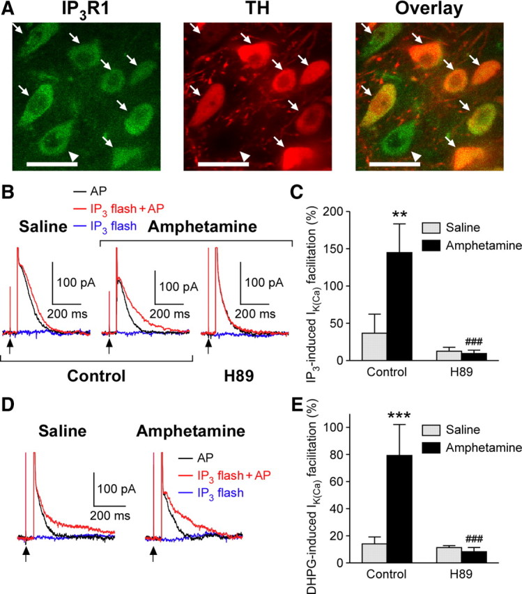
Synaptic plasticity in the mesolimbic dopamine (DA) system is critically involved in reward-based conditioning and the development of drug addiction. Ca2+ signals triggered by postsynaptic action potentials (APs) drive the induction of synaptic plasticity in the CNS. However, it is not clear how AP-evoked Ca2+ signals and the resulting synaptic plasticity are altered during in vivo exposure to drugs of abuse. We have recently described long-term potentiation (LTP) of NMDA receptor (NMDAR)-mediated transmission onto DA neurons that is induced in a manner dependent on bursts of APs. LTP induction requires amplification of burst-evoked Ca2+ signals by preceding activation of metabotropic glutamate receptors (mGluRs) generating inositol 1,4,5-trisphosphate (IP3). In this study, using brain slices prepared from male rats, we show that repeated in vivo exposure to the psychostimulant amphetamine (5 mg/kg, i.p., 3-7 d) upregulates mGluR-dependent facilitation of burst-evoked Ca2+ signals in DA neurons of the ventral tegmental area (VTA). Protein kinase A (PKA)-induced sensitization of IP3 receptors mediates this upregulation of mGluR action. As a consequence, NMDAR-mediated transmission becomes more susceptible to LTP induction after repeated amphetamine exposure. We have also found that the magnitude of amphetamine-conditioned place preference (CPP) in behaving rats correlates with the magnitude of mGluR-dependent Ca2+ signal facilitation measured in VTA slices prepared from these rats. Furthermore, the development of amphetamine CPP is significantly attenuated by intra-VTA infusion of the PKA inhibitor H89. We propose that enhancement of mGluR-dependent NMDAR plasticity in the VTA may promote the learning of environmental stimuli repeatedly associated with amphetamine experience.


Figures

Figure 1.
Figure 1.
APs are responsible for the generation of large AHPs. Representative traces of spontaneous DA neuron firing, recorded with a perforated-patch configuration before and during bath application of TTX (50 nm), are shown. APs can be occasionally observed after brief TTX application (3 min). Note that the amplitude of AHPs after APs is significantly larger than the amplitude of hyperpolarizations during subthreshold membrane potential oscillations. APs were completely eliminated after prolonged perfusion of TTX (9 min).
Figure 2.
Figure 2.
mGluR-dependent facilitation of AP-evoked Ca2+ signals is augmented after withdrawal from repeated, but not single, amphetamine exposure. A, Example traces of IK(Ca) (top) and summary time graphs (bottom) illustrating the effects of DHPG (1 μm) on single AP- and burst-evoked IK(Ca) in VTA DA neurons from saline- and amphetamine-treated rats. B, Summary bar graphs demonstrating that in vivo amphetamine exposure augmented DHPG-induced facilitation of IK(Ca) in the VTA (naïve, n = 5 from 3 rats; saline, n = 14 from 12 rats; amphetamine, n = 18 from 17 rats) but not in the SNc (naïve, n = 5 from 4 rats; saline, n = 9 from 7 rats; amphetamine, n = 10 from 8 rats). *p < 0.05; **p < 0.01; ***p < 0.001. C, A summary bar graph showing that the size of baseline IK(Ca) was not affected by in vivo amphetamine treatment in VTA DA neurons. The time integral of IK(Ca) (expressed in picocoulombs) was normalized to the membrane capacitance in each neuron to evaluate the size of baseline IK(Ca). The numbers of neurons per rats are the same as those for VTA data in B. D, Left, Confocal fluorescence image of a VTA DA neuron filled with Fluo-5F (25 μm) from an amphetamine-treated rat. Fluorescence changes were measured at the ROI covering an area in the proximal dendrite that extended ∼10 μm from the soma. Scale bar, 20 μm. Right, Representative traces illustrating the effect of DHPG on burst-induced Ca2+ signals in saline- and amphetamine-treated rats. Bursts of five APs were elicited at the times indicated. Traces from an amphetamine-treated rat were obtained from the neuron shown on the left. E, Summary graph showing the magnitude of DHPG-induced facilitation of IK(Ca) (single AP) after various amphetamine treatment regimens as indicated (saline, n = 14 from 12 rats; 1 d amphetamine, n = 11 from 7 rats; 3 d amphetamine, n = 5 from 4 rats; 7 d amphetamine, n = 18 from 17 rats; 7 d amphetamine plus 10 d withdrawal, n = 9 from 8 rats). F, Repeated amphetamine exposure did not affect DHPG-induced inward currents. The numbers of neurons per rats are the same as those for VTA data in B. Error bars indicate SEM.
Figure 3.
Figure 3.
IP3R sensitivity is increased after repeated amphetamine exposure. A, Traces of IIP3 evoked with different UV pulse intensities (50, 150, 450, 1350, and 4050 μF) in a VTA DA neuron from an amphetamine-treated rat. The cytosol was loaded with caged IP3 (100 μm). B, Averaged concentration (UV pulse intensity)–response (IIP3) curves in VTA neurons from saline- and amphetamine-treated rats (saline, n = 9 from 6 rats; amphetamine, n = 10 from 8 rats). Data are fitted to a logistic equation. The IIP3 amplitude is normalized to the maximal value. *p < 0.05; **p < 0.01 versus saline group. Error bars indicate SEM.
Figure 4.
Figure 4.
Involvement of PKA in IP3R sensitization after in vivo amphetamine exposure. A, Confocal photomicrographs showing coexpression of IP3R1 (green; left) and TH (red; middle) in VTA neurons. Both images are superimposed to better illustrate cellular colocalization of IP3R1 and TH immunoreactivities (right). Arrows indicate cells coexpressing IP3R1 and TH, whereas the cell with an arrowhead expressed IP3R1 but not TH. Scale bars, 20 μm. B, Representative traces of IK(Ca) (single AP) with and without flash photolysis of caged IP3 (25 μm) in VTA neurons from saline- and amphetamine-treated rats. A low-intensity UV pulse (100 μF) was applied 50 ms before the 2 ms depolarization. Current traces observed with the UV pulse alone without the following depolarization are also shown (blue). Note that IP3-induced facilitation of IK(Ca) observed in amphetamine-treated rats is blocked by H89 (10 μm; right traces). C, Summary bar graph showing the magnitude of IP3-induced facilitation of IK(Ca) under control recording conditions (saline, n = 7 from 3 rats; amphetamine, n = 8 from 6 rats) and in H89 (saline, n = 9 from 4 rats; amphetamine, n = 8 from 4 rats). **p < 0.01 versus saline group; ###p < 0.001 versus control condition. D, Representative traces illustrating that IP3-induced facilitation of IK(Ca) can be observed in the presence of H89 when stronger UV pulses are used (200 μF in the experiments shown). E, Summary bar graph demonstrating the DHPG effect on IK(Ca) under control recording conditions (saline, n = 6 from 5 rats; amphetamine, n = 5 from 5 rats) and in H89 (saline, n = 9 from 7 rats; amphetamine, n = 6 from 4 rats). ***p < 0.001 versus saline group; ###p < 0.001 versus control condition. Error bars indicate SEM.
Figure 5.
Figure 5.
Synaptic facilitation of IK(Ca) is enhanced after repeated amphetamine exposure. A, Representative traces illustrating the difference in synaptic facilitation of IK(Ca) between saline- and amphetamine-treated rats using synaptic stimulation trains of various durations (0.25, 0.5, 1.0, and 1.5 s). A single AP was evoked 100 ms after the offset of each synaptic stimulation train. Traces of IK(Ca) after synaptic stimulation are shown after subtracting the trace elicited by synaptic stimulation alone. All traces obtained with different synaptic stimulation durations are overlaid. B, C, Summary graphs plotting the magnitude of synaptic facilitation of IK(Ca) versus stimulation duration for single APs (saline, n = 7 from 3 rats; amphetamine, n = 5 from 4 rats) (B) and bursts (saline, n = 6 from 3 rats; amphetamine, n = 3 from 3 rats) (C). *p < 0.05; **p < 0.01; ***p < 0.001 versus saline group. Stim, stimulation. Error bars indicate SEM.
Figure 6.
Figure 6.
NMDAR-mediated transmission onto VTA DA neurons is more susceptible to LTP induction by synaptic stimulation–burst pairing in amphetamine-treated rats. A, Example experiments to induce NMDAR LTP in saline- and amphetamine-treated rats. Time graphs of NMDAR EPSC amplitude are shown on the left. The LTP induction protocol, which consisted of repetitive (10 times every 20 s) synaptic stimulation–burst pairing (top right), was delivered at the time indicated by the arrow. Traces of NMDAR EPSCs at times indicated by numbers in the time graphs are also shown. B, Summary time graph of NMDAR LTP experiments performed under control recording conditions from saline- and amphetamine-treated rats and in H89 from amphetamine-treated rats (saline, n = 6 from 4 rats; amphetamine, n = 5 from 4 rats; amphetamine–H89, n = 5 from 5 rats). Each symbol represents mean normalized EPSC amplitude from a 2 min window. Error bars indicate SEM. C, The magnitude of NMDAR LTP is plotted versus the magnitude of synaptic facilitation of IK(Ca) in neurons examined for NMDAR LTP in B. The solid line is a linear fit to all data points.
Figure 7.
Figure 7.
Amphetamine-induced CPP correlates with DHPG-induced facilitation of IK(Ca) measured in brain slices. A, The preference for the amphetamine-paired side during the pretest and the posttest are plotted in 11 rats. ***p < 0.001. B, The magnitude of CPP is plotted versus the magnitude of DHPG-induced facilitation of IK(Ca) for both single APs and bursts. Solid lines represent linear fit to the data.
Figure 8.
Figure 8.
PKA blockade in the VTA attenuates the acquisition of amphetamine CPP. A, Left, Representative photomicrograph of a cresyl violet-stained section illustrating bilateral cannulae placements. This section was obtained from a rat that was injected with H89 during amphetamine conditioning. Arrowheads indicate the tips of injection cannulae. Right, Schematic diagram depicting the approximate locations of cannulae tips in 18 rats from which the data in B and C were obtained. The number on each panel represents the distance (in millimeters) from bregma as indicated by Paxinos and Watson (1998). B, A graph demonstrating that intra-VTA injection of H89 does not affect side preference. Both compartments were paired with intraperitoneal injection of saline in these six rats. C, Changes in the preference for the amphetamine-paired side are shown for rats that received intra-VTA injection of saline (left; 6 rats) or H89 (right; 6 rats) before each amphetamine conditioning session. *p < 0.05; **p < 0.01.
Figure 9.
Figure 9.
Schematic diagram illustrating the NMDAR plasticity mechanism and the effect of repeated amphetamine exposure. Sustained stimulation of glutamatergic inputs produces a gradual increase in cytosolic IP3 levels via activation of mGluRs. Burst-evoked Ca2+ signals, triggered by Ca2+ influx through voltage-gated Ca2+ channels (VGCCs), are amplified if the burst occurs when IP3 levels are elevated. These amplified burst-evoked Ca2+ signals drive the induction of NMDAR LTP. After repeated amphetamine exposure, PKA-mediated phosphorylation of IP3Rs will be upregulated, causing an increase in IP3 sensitivity of IP3Rs. This will promote the induction of NMDAR LTP in amphetamine-treated rats.

Similar articles

Cited by

References

    1. Abraham WC. Metaplasticity: tuning synapses and networks for plasticity. Nat Rev Neurosci. 2008;9:387. - PubMed
    1. Beeler JA, Cao ZF, Kheirbek MA, Zhuang X. Loss of cocaine locomotor response in Pitx3-deficient mice lacking a nigrostriatal pathway. Neuropsychopharmacology. 2009;34:1149–1161. - PMC - PubMed
    1. Bellone C, Luscher C. Cocaine triggered AMPA receptor redistribution is reversed in vivo by mGluR-dependent long-term depression. Nat Neurosci. 2006;9:636–641. - PubMed
    1. Berridge KC. The debate over dopamine's role in reward: the case for incentive salience. Psychopharmacology (Berl) 2007;191:391–431. - PubMed
    1. Bonci A, Williams JT. Increased probability of GABA release during withdrawal from morphine. J Neurosci. 1997;17:796–803. - PMC - PubMed

Publication types

MeSH terms