Muscarinic signaling in the cochlea: presynaptic and postsynaptic effects on efferent feedback and afferent excitability
- PMID: 20463237
- PMCID: PMC3332094
- DOI: 10.1523/JNEUROSCI.5080-09.2010
Muscarinic signaling in the cochlea: presynaptic and postsynaptic effects on efferent feedback and afferent excitability
Abstract
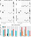
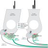
Acetylcholine is the major neurotransmitter of the olivocochlear efferent system, which provides feedback to cochlear hair cells and sensory neurons. To study the role of cochlear muscarinic receptors, we studied receptor localization with immunohistochemistry and reverse transcription-PCR and measured olivocochlear function, cochlear responses, and histopathology in mice with targeted deletion of each of the five receptor subtypes. M2, M4, and M5 were detected in microdissected immature (postnatal days 10-13) inner hair cells and spiral ganglion cells but not outer hair cells. In the adult (6 weeks), the same transcripts were found in microdissected organ of Corti and spiral ganglion samples. M2 protein was found, by immunohistochemistry, in olivocochlear fibers in both outer and inner hair cell areas. M3 mRNA was amplified only from whole cochleas, and M1 message was never seen in wild-type ears. Auditory brainstem responses (ABRs) and distortion product otoacoustic emissions (DPOAEs) were unaffected by loss of Gq-coupled receptors (M1, M3, or M5), as were shock-evoked olivocochlear effects and vulnerability to acoustic injury. In contrast, loss of Gi-coupled receptors (M2 and/or M4) decreased neural responses without affecting DPOAEs (at low frequencies). This phenotype and the expression pattern are consistent with excitatory muscarinic signaling in cochlear sensory neurons. At high frequencies, both ABRs and DPOAEs were attenuated by loss of M2 and/or M4, and the vulnerability to acoustic injury was dramatically decreased. This aspect of the phenotype and the expression pattern are consistent with a presynaptic role for muscarinic autoreceptors in decreasing ACh release from olivocochlear terminals during high-level acoustic stimulation and suggest that muscarinic antagonists could enhance the resistance of the inner ear to noise-induced hearing loss.
Figures


Similar articles
-
Dopaminergic signaling in the cochlea: receptor expression patterns and deletion phenotypes.J Neurosci. 2012 Jan 4;32(1):344-55. doi: 10.1523/JNEUROSCI.4720-11.2012. J Neurosci. 2012. PMID: 22219295 Free PMC article.
-
Loss of GABAB receptors in cochlear neurons: threshold elevation suggests modulation of outer hair cell function by type II afferent fibers.J Assoc Res Otolaryngol. 2009 Mar;10(1):50-63. doi: 10.1007/s10162-008-0138-7. Epub 2008 Oct 17. J Assoc Res Otolaryngol. 2009. PMID: 18925381 Free PMC article.
-
Muscarinic receptor subtypes are differentially distributed in the rat cochlea.Neuroscience. 2002;111(2):291-302. doi: 10.1016/s0306-4522(02)00020-9. Neuroscience. 2002. PMID: 11983315
-
The afferent signaling complex: Regulation of type I spiral ganglion neuron responses in the auditory periphery.Hear Res. 2016 Jun;336:1-16. doi: 10.1016/j.heares.2016.03.011. Epub 2016 Mar 25. Hear Res. 2016. PMID: 27018296 Review.
-
Efferent Inhibition of the Cochlea.Cold Spring Harb Perspect Med. 2019 May 1;9(5):a033530. doi: 10.1101/cshperspect.a033530. Cold Spring Harb Perspect Med. 2019. PMID: 30082454 Free PMC article. Review.
Cited by
-
Bile Acid Application in Cell-Targeting for Molecular Receptors in Relation to Hearing: A Comprehensive Review.Curr Drug Targets. 2024;25(3):158-170. doi: 10.2174/0113894501278292231223035733. Curr Drug Targets. 2024. PMID: 38192136 Review.
-
Contralateral-noise effects on cochlear responses in anesthetized mice are dominated by feedback from an unknown pathway.J Neurophysiol. 2012 Jul;108(2):491-500. doi: 10.1152/jn.01050.2011. Epub 2012 Apr 18. J Neurophysiol. 2012. PMID: 22514298 Free PMC article.
-
Disruption of lateral olivocochlear neurons with a dopaminergic neurotoxin depresses spontaneous auditory nerve activity.Neurosci Lett. 2014 Oct 17;582:54-8. doi: 10.1016/j.neulet.2014.08.040. Epub 2014 Aug 29. Neurosci Lett. 2014. PMID: 25175420 Free PMC article.
-
Sound exposure dynamically induces dopamine synthesis in cholinergic LOC efferents for feedback to auditory nerve fibers.Elife. 2020 Jan 24;9:e52419. doi: 10.7554/eLife.52419. Elife. 2020. PMID: 31975688 Free PMC article.
-
Characterization of the transcriptome of nascent hair cells and identification of direct targets of the Atoh1 transcription factor.J Neurosci. 2015 Apr 8;35(14):5870-83. doi: 10.1523/JNEUROSCI.5083-14.2015. J Neurosci. 2015. PMID: 25855195 Free PMC article.
References
-
- Bartolami S, Ripoll C, Planche M, Pujol R. Localization of functional muscarinic receptors in the rat cochlea: evidence for efferent presynaptic autoreceptors. Brain Res. 1993;626:200–209. - PubMed
-
- Brown DA, Sihra TS. Presynaptic signaling by heterotrimeric G-proteins. Handb Exp Pharmacol. 2008;184:207–260. - PubMed
-
- Caulfield MP. Muscarinic receptors-characterization, coupling and function. Pharmacol Ther. 1993;58:319–379. - PubMed
-
- Chen Z, Kujawa SG, Sewell WF. Auditory sensitivity regulation via rapid changes in expression of surface AMPA receptors. Nat Neurosci. 2007;10:1238–1240. - PubMed
Publication types
MeSH terms
Substances
Grants and funding
LinkOut - more resources
Full Text Sources
Other Literature Sources
Molecular Biology Databases
