. 2010 May 12;30(19):6763-75.
doi: 10.1523/JNEUROSCI.0801-10.2010.

Dicer in Schwann cells is required for myelination and axonal integrity

Affiliations

Dicer in Schwann cells is required for myelination and axonal integrity

Jorge A Pereira et al. J Neurosci. .

Abstract

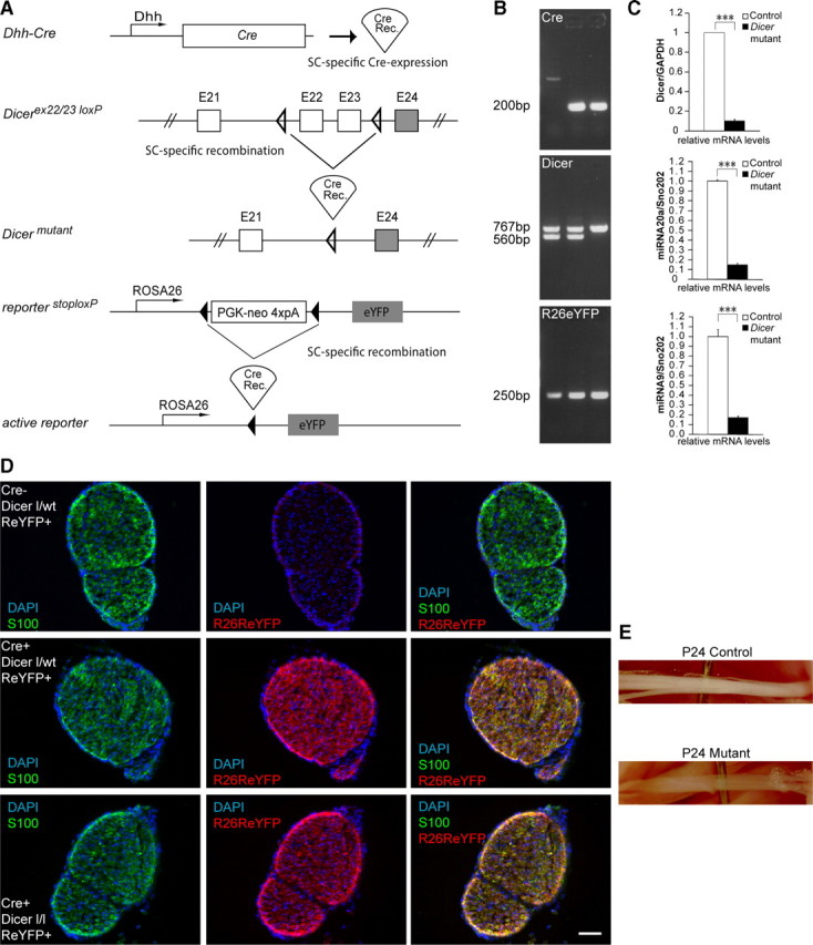
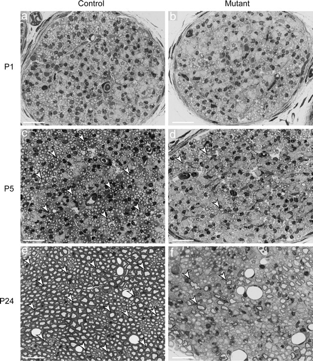
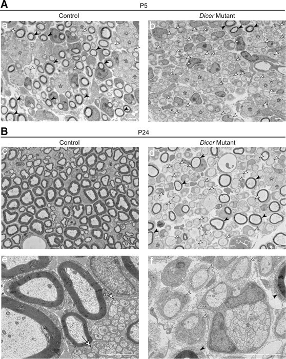
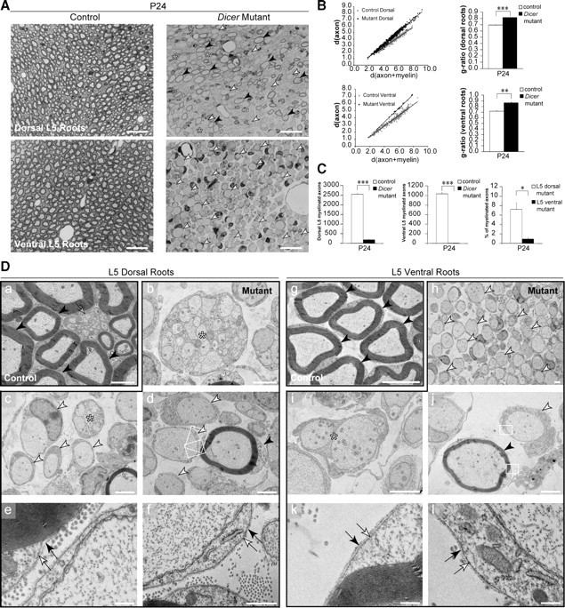
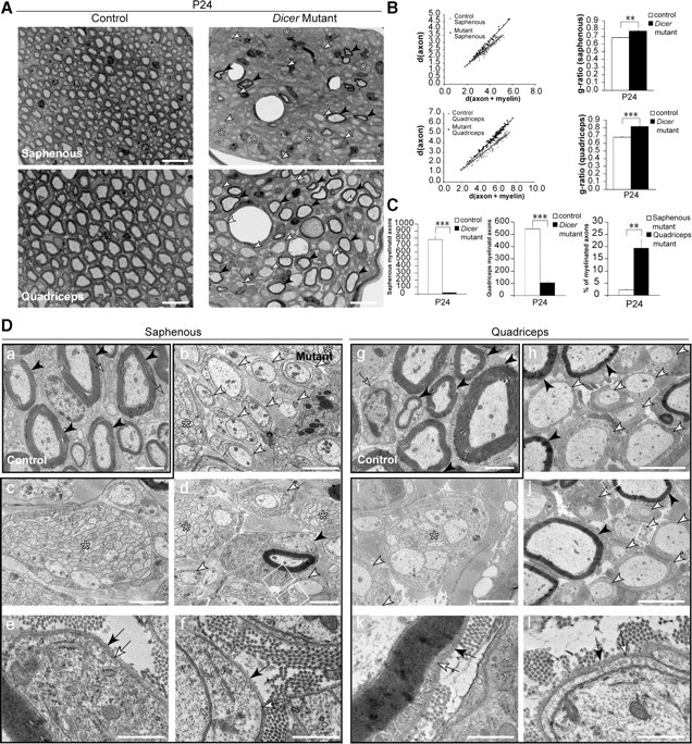
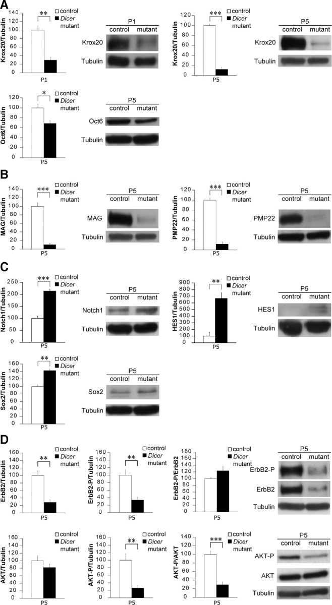
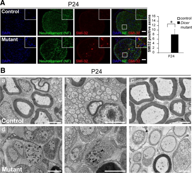
Dicer is responsible for the generation of mature micro-RNAs (miRNAs) and loading them into RNA-induced silencing complex (RISC). RISC functions as a probe that targets mRNAs leading to translational suppression and mRNA degradation. Schwann cells (SCs) in the peripheral nervous system undergo remarkable differentiation both in morphology and gene expression patterns throughout lineage progression to myelinating and nonmyelinating phenotypes. Gene expression in SCs is particularly tightly regulated and critical for the organism, as highlighted by the fact that a 50% decrease or an increase to 150% of normal gene expression of some myelin proteins, like PMP22, results in peripheral neuropathies. Here, we selectively deleted Dicer and consequently gene expression regulation by mature miRNAs from Mus musculus SCs. Our results show that in the absence of Dicer, most SCs arrest at the promyelinating stage and fail to start forming myelin. At the molecular level, the promyelinating transcription factor Krox20 and several myelin proteins [including myelin associated glycoprotein (MAG) and PMP22] were strongly reduced in mutant sciatic nerves. In contrast, the myelination inhibitors SOX2, Notch1, and Hes1 were increased, providing an additional potential basis for impaired myelination. A minor fraction of SCs, with some peculiar differences between sensory and motor fibers, overcame the myelination block and formed unusually thin myelin, in line with observed impaired neuregulin and AKT signaling. Surprisingly, we also found signs of axonal degeneration in Dicer mutant mice. Thus, our data indicate that miRNAs critically regulate Schwann cell gene expression that is required for myelination and to maintain axons via axon-glia interactions.


Figures

Figure 1.
Figure 1.
Recombination of the conditional Dicer allele in Schwann cells of mutant mice. A, Regulatory elements of the Dhh promoter drive the expression of Cre in SCs. Schematic map of the Dicer allele depicts the location of the loxP sequences. On Cre-mediated recombination, the genomic region located between the two loxP sites, which includes exon 22 and 23, is excised, thereby inactivating the conditional Dicer allele. The Rosa26 stop/flox locus annexed to an eYFP reporter construct was bred into the floxed animals. When cre recombinase is expressed in SCs, it excises the stop cassette and allows expression of eYFP. B, PCR performed on genomic DNA using primers against Cre (top; 200 bp), Dicer (middle; floxed 767 bp, wt 560 bp), and R26R locus (bottom; 250 bp) showed a cre-negative Dicer flox/wt animal in lane 1 (control), cre-positive Dicer flox/wt animal in lane 2 (heterozygous), and a cre-positive Dicer flox/flox animal in lane 3 (mutant). All three animals were carrying the R26R eYFP construct. C, qRT-PCR performed on RNA extracted from three independent P5 animals showed a dramatic reduction of Dicer (top) mRNA in mutant animals (n = 3, p < 0.0001). The low residual Dicer mRNA levels detected in mutant lysates are likely the result of the presence of endoneurial fibroblasts. qRT-PCR targeting mature miRNAs 20a (middle) (n = 3, p = 0.00031) and 9 (bottom) (n = 3, p = 0.00032) revealed a dramatic decrease of mature miRNAs in Dicer mutant SNs at P5. D, Immunohistochemistry performed on SNs from P3 animals using antibodies against S100 (green channel) and Rosa26 eYFP (red channel). Animals not expressing Cre showed no signal for Rosa26 eYFP (top), whereas control (middle) or mutant mice (bottom) carrying the Cre and Rosa26 reporter displayed an abundant signal for the eYFP antibody, colocalizing with S100, indicating that recombination successfully occurred in the SCs of these animals. E, Control and mutant SNs from P24 littermates. Note that SNs of mutant mice were thinner and more transparent than controls. Error bars display ± SEM. Scale bars, 50 μm.
Figure 2.
Figure 2.
Myelination is impaired on loss of Dicer in sciatic nerves. a–f, SN cross sections (0.5 μm) stained with toluidine blue. a–d, Early in development, SCs are in close association with bundles of axons (asterisks). Progressively, individual axons are sorted from these bundles in a process referred to as radial sorting. E, In control nerves, individual large-caliber axons were wrapped and myelinated. f, In the mutant nerves, myelination was impaired, and only a few myelinated axons were visible at P24 (arrowheads), together with a few small bundles of unsorted axons (asterisks) and many 1:1 relations between axons and SCs, which did not present myelin. Scale bars, 20 μm.
Figure 3.
Figure 3.
Schwann cells were mainly arrested at the promyelinating stage in the absence of mature miRNAs. A, B, EM micrographs of SNs ultrathin sections at P5 (A) and P24 (B). A, At P5, a panoramic merge of several high-magnification photographs showed in controls (a) a mixture of unsorted axonal bundles (asterisks), 1:1 relations that were not yet myelinated (white arrowheads), and numerous myelinated figures (black arrowheads). Mutant sciatic nerves (b) also displayed several bundles (asterisks), but very few myelinated fibers (black arrowheads) and many 1:1 not-myelinated profiles (white arrowheads). B, At P24, control SNs were extensively myelinated (c, e) and nonmyelinating SCs had established close relations with very small-caliber axons (e, white arrows). In Dicer mutants, a panoramic photomerge (d) showed abundant 1:1 not-myelinated relations (d, f, white arrowheads), some remaining unsorted bundles of naked axons (d, f, asterisks) and a few fibers that had been successfully myelinated (d, f, black arrowheads). Scale bars, 2 μm.
Figure 4.
Figure 4.
Dorsal and ventral roots were distinctively affected on loss of Dicer in Schwann cells. A, Toluidine blue-stained sections (0.5 μm) of P24 dorsal (sensory fiber-rich) and ventral (motor fiber-rich) roots showed widespread myelination in control animals, opposed to impaired myelination in the mutants. Mutant dorsal roots presented few myelinated fibers (black arrowheads) and small bundles of unsorted axons (asterisks). Dorsal and ventral mutant roots displayed many 1:1 not-myelinated profiles (white arrowheads). Scale bars, 20 μm. B, Scatterplot graphics of measurements performed in control and mutant animals showed thinner myelin sheaths on fibers that managed to get myelinated in mutant animals, both in dorsal and ventral roots. Thinner myelin sheaths were translated into higher values for g-ratio in every animal analyzed (n = 3, p < 0.0001 for dorsal roots, and n = 3, p = 0.0013 for ventral roots). C, The total number of myelinated fibers was quantified in the L5 roots of control and mutant animals. Both dorsal (n = 3, p < 0.0001) and ventral (n = 3, p < 0.0001) roots displayed a small fraction of myelinated fibers when compared with control counterparts. Interestingly, when comparing mutant dorsal to mutant ventral roots we observed that the percentage of myelinated fibers in dorsal roots was about eightfold higher (n = 3, p = 0.011). Da–l, EM micrographs of dorsal roots (a–f) and ventral roots (g–l). Control roots (a, g) displayed thoroughly myelinated fibers (black arrowheads), and in control dorsal roots we could also find nonmyelinating SCs (gray arrow). Mutant dorsal roots displayed bundles of naked axons (b, c, asterisks), 1:1 not-myelinated profiles (c, d, white arrowheads), and some myelinated fibers (d, black arrowhead). Mutant ventral roots were filled with 1:1 not-myelinated profiles (h, white arrowheads), and sporadically SCs were engaging more than a single axon (i, asterisk), although these structures were different from the bundles observed in the dorsal roots. The myelinating cells and the 1:1 profiles in the roots displayed a basal lamina (e, f, k, l, black arrows) apposed to the plasmalemma (e, f, k, l, white arrows), indicating that these were SCs and not invading oligodendrocytes. Scale bars: g, 5 μm; a–d, h–j, 2 μm; e, 0.5 μm; f, k, l, 0.2 μm. Error bars display ± SEM.
Figure 5.
Figure 5.
Sensory- and motor-enriched femoralis branches were distinctively affected on loss of Dicer in Schwann cells. A, Toluidine blue-stained sections (0.5 μm) of P24 saphenous nerve, exclusively composed of sensory axons, and quadriceps nerve, composed of 40–50% myelinated motor axons. Control nerves were widely myelinated, but mutant nerves displayed very few myelinated fibers (black arrowheads), several promyelinating profiles (white arrowheads), and some small axonal bundles (asterisks). Scale bars, 20 μm. B, Scatterplot graphics of measurements performed in control and mutant animals showed thinner myelin sheaths on fibers that were myelinated in mutant animals. Every mutant animal analyzed displayed a higher average g-ratio than control littermates (n = 3, p = 0.0014 for dorsal roots; n = 3, p < 0.0001 for ventral roots). C, The total number of myelinated fibers was quantified in the entire nerve cross section of control and mutant animals. Both saphenous (n = 3, p = 0.00011) and quadriceps (n = 3, p < 0.0001) nerves displayed a small fraction of myelinated fibers when compared with control counterparts. Surprisingly, when comparing myelinated fibers in the mutant saphenous nerves to the mutant quadriceps nerves, the saphenous nerve showed an approximately ninefold lower percentage of myelinated axons than the quadriceps (n = 3, p = 0.0077). Da–l, EM micrographs of saphenous (a–f) and quadriceps (g–l) nerves. Control nerves (a, g) displayed thoroughly myelinated fibers (black arrowheads) and mature nonmyelinating SCs (gray arrows). Mutant saphenous nerves displayed bundles of naked axons (b–d, asterisks), 1:1 not-myelinated profiles (b, d, white arrowheads), and very rare myelinated fibers (d, black arrowhead). Quadriceps nerves from Dicer mutants presented 1:1 not-myelinated profiles (h–j, white arrowheads), bundles of unsorted axons (i, j, asterisks), and some myelinating SCs (h, j, black arrowheads). The myelinating and promyelinating SCs presented a basal lamina (e, f, k, l, black arrow) tightly apposed to the SC abaxonal plasmalemma (e, f, k, l, white arrow). Both mutant nerves displayed very high levels of collagen when compared with controls. Scale bars: h, j, 5 μm; a–d, g, i, 2 μm; e, f, k, l, 0.5 μm. Error bars display ± SEM.
Figure 6.
Figure 6.
Loss of Dicer resulted in failure to downregulate myelination inhibitors and impaired myelin-stimulating pathways in mouse sciatic nerves. A, Krox20 protein levels were heavily reduced at P5 (n = 5, p < 0.0001) and even as early as P1 (n = 3, p = 0.0045) in mutant Dicer SN lysates. Oct6 levels were also slightly reduced in mutants at P5 (n = 3, p = 0.031). B, The myelin proteins MAG (n = 5, p < 0.0001) and PMP22 (n = 3, p = 0.00017) were also reduced in Dicer mutant mice. C, The transcription factor SOX2, an inhibitor of myelination, was increased in mutant SNs at P5 compared with controls (n = 3, p = 0.0022). NICD was present at higher levels in mutants (n = 3, p = 0.00068), as was its downstream effector HES1 (n = 3, p = 0.0049), which can also act as myelination inhibitor. D, The neuregulin receptor ErbB2 in SCs was decreased at the total protein level (n = 7, p = 0.0018), which is reflected in a reduction of the phosphorylated form (tyrosine 1248) when normalized to tubulin (n = 7, p = 0.0021). AKT phosphorylation on serine 473 was also reduced in P5 mutant nerves when compared with total AKT (n = 6, p < 0.0001) or when compared with tubulin (n = 6, p = 0.0029). AKT total levels were unchanged between control and mutant SNs. Error bars display ± SEM.
Figure 7.
Figure 7.
Axonal degeneration on conditional ablation of Dicer in Schwann cells. A, Immunohistochemistry in P24 control and mutant SNs using antibodies against neurofilament (green channel) and SMI32, which specifically recognizes nonphosphorylated neurofilament H (red channel). Zoom boxes highlight the colocalization of the SMI32 and the NF signals. Nerves obtained from Dicer mutant animals showed an SMI32-positive signal in every cross section analyzed, whereas control animals did not show a single positive signal (three different control and mutant animals were analyzed, four full cross sections per animal). Quantification depicts SMI32-positive signal count per section (n = 3, p = 0.026). Scale bars, 50 μm. Error bars display ± SEM. B, Electron micrographs of P24 control (a–c) and mutant (d–f) SNs. In controls, axons that were either myelinated or engaged by a nonmyelinating SC displayed sporadic organelles at the cross-section level (a–c); however, in the mutant animals there were several axons that presented an unusually high accumulation of vesicular structures (d–f, black arrowheads), an indication that the axon might be affected. Scale bars, 2 μm.

Comment in

Similar articles

Cited by

References

    1. Amaral PP, Dinger ME, Mercer TR, Mattick JS. The eukaryotic genome as an RNA machine. Science. 2008;319:1787–1789. - PubMed
    1. Bernstein E, Kim SY, Carmell MA, Murchison EP, Alcorn H, Li MZ, Mills AA, Elledge SJ, Anderson KV, Hannon GJ. Dicer is essential for mouse development. Nat Genet. 2003;35:215–217. - PubMed
    1. Britsch S, Li L, Kirchhoff S, Theuring F, Brinkmann V, Birchmeier C, Riethmacher D. The ErbB2 and ErbB3 receptors and their ligand, neuregulin-1, are essential for development of the sympathetic nervous system. Genes Dev. 1998;12:1825–1836. - PMC - PubMed
    1. Bunge RP. Expanding roles for the Schwann cell: ensheathment, myelination, trophism and regeneration. Curr Opin Neurobiol. 1993;3:805–809. - PubMed
    1. Campana WM, Darin SJ, O'Brien JS. Phosphatidylinositol 3-kinase and Akt protein kinase mediate IGF-I- and prosaptide-induced survival in Schwann cells. J Neurosci Res. 1999;57:332–341. - PubMed

Publication types

MeSH terms