Enteric glia are targets of the sympathetic innervation of the myenteric plexus in the guinea pig distal colon
- PMID: 20463242
- PMCID: PMC6632550
- DOI: 10.1523/JNEUROSCI.0603-10.2010
Enteric glia are targets of the sympathetic innervation of the myenteric plexus in the guinea pig distal colon
Abstract
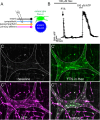
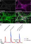
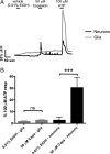
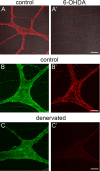
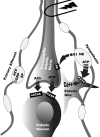
Astrocytes respond to synaptic activity in the CNS. Astrocytic responses are synapse specific and precisely regulate synaptic activity. Glia in the peripheral nervous system also respond to neuronal activity, but it is unknown whether glial responses are synapse specific. We addressed this issue by examining the activation of enteric glia by distinct neuronal subpopulations in the enteric nervous system. Enteric glia are unique peripheral glia that surround enteric neurons and respond to neuronally released ATP with increases in intracellular calcium ([Ca2+]i). Autonomic control of colonic function is mediated by intrinsic (enteric) and extrinsic (sympathetic, parasympathetic, primary afferent) neural pathways. Here we test the hypothesis that a defined population of neurons activates enteric glia using a variety of techniques to ablate or stimulate components of the autonomic innervation of the colon. Our findings demonstrate that, in the male guinea pig colon, activation of intrinsic neurons does not stimulate glial [Ca2+]i responses and fast enteric neurotransmission is not necessary to initiate glial responses. However, ablating extrinsic innervation significantly reduces glial responses to neuronal activation. Activation of primary afferent fibers does not activate glial [Ca2+]i responses. Selectively ablating sympathetic fibers reduces glial activation to a similar extent as total extrinsic denervation. Neuronal activation of glia follows the same frequency dependence as sympathetic neurotransmitter release, but the only sympathetic neurotransmitter that activates glial [Ca2+]i responses is ATP, suggesting that sympathetic fibers release ATP to activate enteric glia. Therefore, enteric glia discern activity in adjacent synaptic pathways and selectively respond to sympathetic activation.
Figures

Similar articles
-
Purinergic neuron-to-glia signaling in the enteric nervous system.Gastroenterology. 2009 Apr;136(4):1349-58. doi: 10.1053/j.gastro.2008.12.058. Epub 2009 Jan 1. Gastroenterology. 2009. PMID: 19250649
-
Functional Intraregional and Interregional Heterogeneity between Myenteric Glial Cells of the Colon and Duodenum in Mice.J Neurosci. 2022 Nov 16;42(46):8694-8708. doi: 10.1523/JNEUROSCI.2379-20.2022. Epub 2022 Nov 1. J Neurosci. 2022. PMID: 36319118 Free PMC article.
-
Targeted electrophysiological analysis of viscerofugal neurons in the myenteric plexus of guinea-pig colon.Neuroscience. 2014 Sep 5;275:272-84. doi: 10.1016/j.neuroscience.2014.04.066. Epub 2014 May 9. Neuroscience. 2014. PMID: 24814020
-
Analysis of enteric neurons, glia and their interactions using explant cultures of the myenteric plexus.Dev Neurosci. 1987;9(4):201-27. doi: 10.1159/000111623. Dev Neurosci. 1987. PMID: 3322784 Review.
-
Purinergic modulation of synaptic signalling at the neuromuscular junction.Pflugers Arch. 2006 Aug;452(5):608-14. doi: 10.1007/s00424-006-0068-3. Epub 2006 Apr 8. Pflugers Arch. 2006. PMID: 16604367 Review.
Cited by
-
Structurally defined signaling in neuro-glia units in the enteric nervous system.Glia. 2019 Jun;67(6):1167-1178. doi: 10.1002/glia.23596. Epub 2019 Feb 7. Glia. 2019. PMID: 30730592 Free PMC article.
-
Macrophages associated with the intrinsic and extrinsic autonomic innervation of the rat gastrointestinal tract.Auton Neurosci. 2012 Jul 2;169(1):12-27. doi: 10.1016/j.autneu.2012.02.004. Epub 2012 Mar 20. Auton Neurosci. 2012. PMID: 22436622 Free PMC article.
-
Mini-review: Intercellular communication between enteric glia and neurons.Neurosci Lett. 2023 May 29;806:137263. doi: 10.1016/j.neulet.2023.137263. Epub 2023 Apr 20. Neurosci Lett. 2023. PMID: 37085112 Free PMC article. Review.
-
Sympathetic Input to Multiple Cell Types in Mouse and Human Colon Produces Region-Specific Responses.Gastroenterology. 2021 Mar;160(4):1208-1223.e4. doi: 10.1053/j.gastro.2020.09.030. Epub 2020 Sep 24. Gastroenterology. 2021. PMID: 32980343 Free PMC article.
-
The purinergic neurotransmitter revisited: a single substance or multiple players?Pharmacol Ther. 2014 Nov;144(2):162-91. doi: 10.1016/j.pharmthera.2014.05.012. Epub 2014 Jun 2. Pharmacol Ther. 2014. PMID: 24887688 Free PMC article. Review.
References
-
- Al-Humayyd M, White TD. Adrenergic and possible nonadrenergic sources of adenosine 5′-triphosphate release from nerve varicosities isolated from ileal myenteric plexus. J Pharmacol Exp Ther. 1985;233:796–800. - PubMed
-
- Blandizzi C, Fornai M, Colucci R, Baschiera F, Barbara G, De Giorgio R, De Ponti F, Breschi MC, Del Tacca M. Altered prejunctional modulation of intestinal cholinergic and noradrenergic pathways by alpha2-adrenoceptors in the presence of experimental colitis. Br J Pharmacol. 2003;139:309–320. - PMC - PubMed
Publication types
MeSH terms
Substances
Grants and funding
LinkOut - more resources
Full Text Sources
Other Literature Sources
Miscellaneous
