Review
. 2010;20 Suppl 2(Suppl 2):S551-68.
doi: 10.3233/JAD-2010-100354.

Mitochondrial iron metabolism and its role in neurodegeneration

Affiliations
Review

Mitochondrial iron metabolism and its role in neurodegeneration

Maxx P Horowitz et al. J Alzheimers Dis. 2010.

Abstract

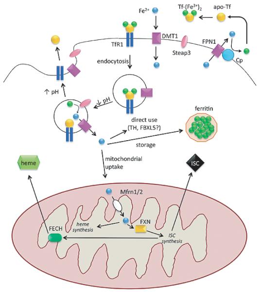
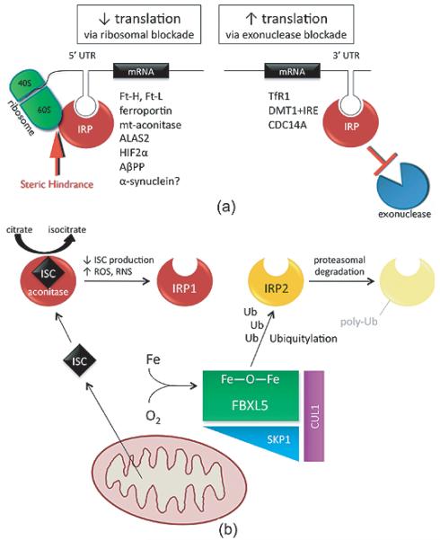
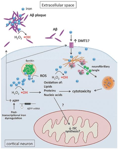
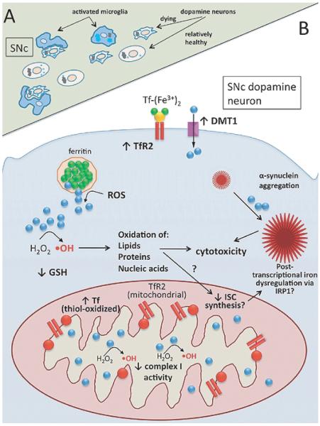
In addition to their well-established role in providing the cell with ATP, mitochondria are the source of iron-sulfur clusters (ISCs) and heme - prosthetic groups that are utilized by proteins throughout the cell in various critical processes. The post-transcriptional system that mammalian cells use to regulate intracellular iron homeostasis depends, in part, upon the synthesis of ISCs in mitochondria. Thus, proper mitochondrial function is crucial to cellular iron homeostasis. Many neurodegenerative diseases are marked by mitochondrial impairment, brain iron accumulation, and oxidative stress - pathologies that are inter-related. This review discusses the physiological role that mitochondria play in cellular iron homeostasis and, in so doing, attempts to clarify how mitochondrial dysfunction may initiate and/or contribute to iron dysregulation in the context of neurodegenerative disease. We review what is currently known about the entry of iron into mitochondria, the ways in which iron is utilized therein, and how mitochondria are integrated into the system of iron homeostasis in mammalian cells. Lastly, we turn to recent advances in our understanding of iron dysregulation in two neurodegenerative diseases (Alzheimer's disease and Parkinson's disease), and discuss the use of iron chelation as a potential therapeutic approach to neurodegenerative disease.


Figures

Fig. 1
Fig. 1
Components of cellular iron homeostasis – import, storage, utilization, export – and their regulation. Endocytotic cycling of the diferric transferrin/transferrin receptor 1 complex and iron release from endosome are depicted in the top center. Upon acidification, ferrous iron released from the endosome may be: 1) used directly as a cofactor for cytosolic proteins; 2) stored in ferritin in its ferric form; or 3) imported into mitochondria. Mitochondria use iron for synthesis of iron-sulfur clusters or heme prosthetic groups, which are then distributed and utilized throughout the cell. The heme synthesis enzyme, ferrochelatase, requires an iron-sulfur cluster for its function. Ferroportin is the only known cellular iron exporter and requires the action of ceruloplasmin to oxidize the exported ferrous iron. Abbreviations: ferrous iron, Fe2+; ferric iron Fe3+; apo-transferrin, apo-Tf; diferric transferrin, Tf-(Fe3+ )2; transferrin receptor 1, TfR1; divalent metal transporter, DMT1; ferroportin, FPN1; ceruloplasmin, Cp; iron-sulfur cluster, ISC; mitoferrin 1/2, Mfrn1/2; frataxin, FXN; ferrochelatase, FECH; tyrosine hydroxylase, TH.
Fig. 2
Fig. 2
Iron regulatory proteins control cellular iron homeostasis. A) Transcripts containing iron-regulatory elements (IREs) in either their 5′ or 3′ untranslated regions (UTR) are bound by iron regulatory proteins (IRP) when iron levels are perceived to be low. IRP binding to 5′ IREs provides steric hindrance to ribosomal machinery, leading to decreased translation of the transcript. IRP binding to 3′ IREs blocks the action of exonucleases, thereby protecting the transcripts from degradation and increasing translation. Transcripts that contain IREs in either their 5′ or 3′ UTRs are listed. Note: There are four isoforms of DMT1 mRNA, only some of which contain 3′ UTR IREs. Abbreviations: ferritin-H/-L, Ft-H/-L; mitochondrial aconitase, mt-aconitase; aminolevulinic acid synthase 2, ALAS2; hypoxia inducible factor 2α, HIF2 α; amyloid β precursor protein, AβPP; transferrin receptor 1, TfR1; divalent metal transporter with IRE, DMT1+IRE; cell division cycle 14 homolog A, CDC14A. B) When it contains a functional ISC, IRP1 (red) functions as a cytosolic aconitase (converting citrate to isocitrate); in the absence of a functional ISC it is converted to an IRE-binding protein. The status of the ISC can change in response to decreased ISC production or increased ISC disruption by reactive oxygen species (ROS). IRP2 (yellow) is a constitutive IRE-binding protein that is regulated by proteasomal degradation. When cellular iron and oxygen levels are adequate, FBXL5 (green) can complex with SKP1 (blue) and CUL1 (purple) to form a functional E3 ubiquitin ligase that tags IRP2 for degradation (light yellow).
Fig. 3
Fig. 3
Iron dysregulation in Alzheimer’s disease. Subcellular iron-related pathology in cortical neurons includes: Fenton chemistry (hydroxyl radical formation, ·OH); interaction of iron (blue) with Aβ (purple) in the extracellular space; iron-induced (green) aggregation of hyper-phosphorylated (pink) tau (black) and neurofibrillary tangle formation; increased expression of DMT1; increased Aβ production, with possible contribution from IRP dysregulation.
Fig. 4
Fig. 4
Iron dysregulation in Parkinson’s disease. A) Tissue-level view of the substantia nigra pars compacta (SNc) showing suspected ante-mortem pathology: iron-laden (light blue circles), degenerating neuromelanin (black) containing neurons surrounded by activated microglia (blue). B) Subcellular view of nigral dopamine neuron showing pathophysiology and suspected mechanisms of iron dysregulation. Key features: decreased complex I activity; α-synuclein aggregation, exacerbated by iron; increased TfR2 and DMT1 protein levels at plasma membrane; expression of TfR2 in nigral mitochondria; accumulation of oxidized Tf (red) in mitochondria – with associated reductive release of ferrous iron (blue); ROS-mediated release of ferrous iron from ferritin; Fenton chemistry (hydroxyl radical formation, ·OH); decreased levels of reduced glutathione (GSH).

References

    1. Herrero-Mendez A, Almeida A, Fernandez E, Maestre C, Moncada S, Bolanos JP. The bioenergetic and antioxidant status of neurons is controlled by continuous degradation of a key glycolytic enzyme by APC/C-Cdh1. Nat Cell Biol. 2009;11:747–752. - PubMed
    1. Sheftel A, Stehling O, Lill R. Iron-sulfur proteins in health and disease. Trends Endocrinol Metab. 2010;21:302–314. - PubMed
    1. Furuyama K, Kaneko K, Vargas PD. Heme as a magnificent molecule with multiple missions: heme determines its own fate and governs cellular homeostasis. Tohoku J Exp Med. 2007;213:1–16. - PubMed
    1. MacKenzie EL, Iwasaki K, Tsuji Y. Intracellular iron transport and storage: from molecular mechanisms to health implications. Antioxid Redox Signal. 2008;10:997–1030. - PMC - PubMed
    1. Garrick MD, Garrick LM. Cellular iron transport. Biochim Biophys Acta. 2009;1790:309–325. - PubMed

Publication types