. 2010;20 Suppl 2(0 2):S293-310.
doi: 10.3233/JAD-2010-100351.

Systemic mitochondrial dysfunction and the etiology of Alzheimer's disease and down syndrome dementia

Affiliations

Systemic mitochondrial dysfunction and the etiology of Alzheimer's disease and down syndrome dementia

Pinar E Coskun et al. J Alzheimers Dis. 2010.

Abstract

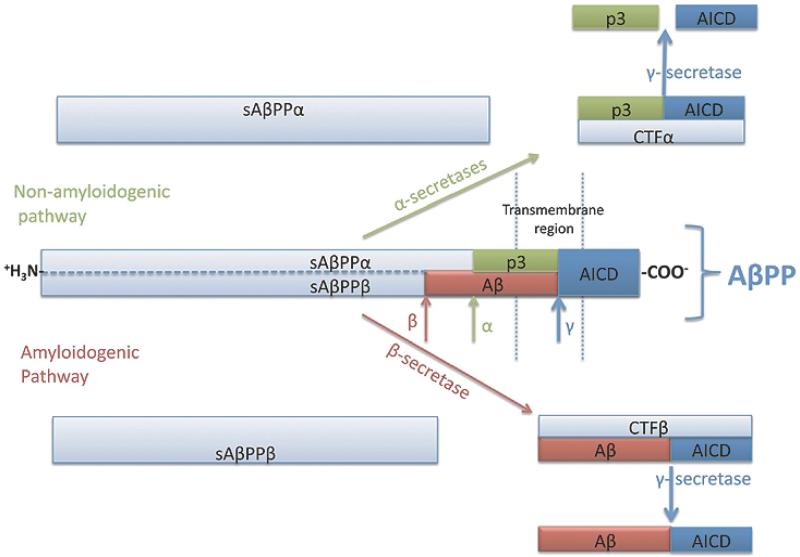
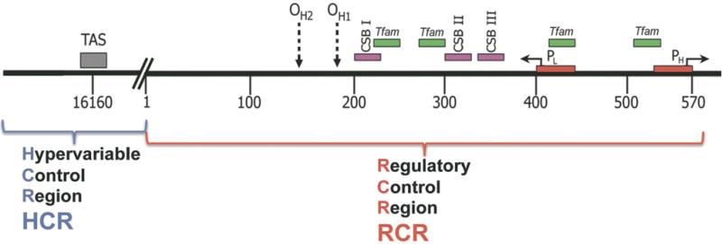
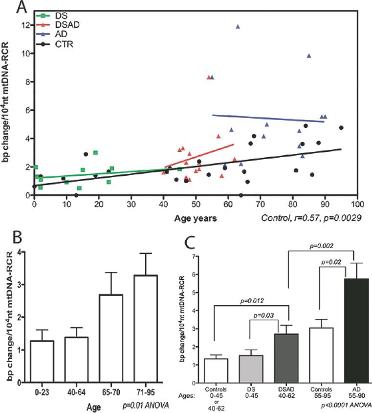
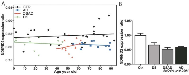
Increasing evidence is implicating mitochondrial dysfunction as a central factor in the etiology of Alzheimer's disease (AD). The most significant risk factor in AD is advanced age and an important neuropathological correlate of AD is the deposition of amyloid-beta peptide (Abeta40 and Abeta42) in the brain. An AD-like dementia is also common in older individuals with Down syndrome (DS), though with a much earlier onset. We have shown that somatic mitochondrial DNA (mtDNA) control region (CR) mutations accumulate with age in post-mitotic tissues including the brain and that the level of mtDNA mutations is markedly elevated in the brains of AD patients. The elevated mtDNA CR mutations in AD brains are associated with a reduction in the mtDNA copy number and in the mtDNA L-strand transcript levels. We now show that mtDNA CR mutations increase with age in control brains; that they are markedly elevated in the brains of AD and DS and dementia (DSAD) patients; and that the increased mtDNA CR mutation rate in DSAD brains is associated with reduced mtDNA copy number and L-strand transcripts. The increased mtDNA CR mutation rate is also seen in peripheral blood DNA and in lymphoblastoid cell DNAs of AD and DSAD patients, and distinctive somatic mtDNA mutations, often at high heteroplasmy levels, are seen in AD and DSAD brain and blood cells DNA. In aging, DS, and DSAD, the mtDNA mutation level is positively correlated with beta-secretase activity and mtDNA copy number is inversely correlated with insoluble Abeta40 and Abeta42 levels. Therefore, mtDNA alterations may be responsible for both age-related dementia and the associated neuropathological changes observed in AD and DSAD.


Figures

Fig. 1
Fig. 1
Schematic representation of AβPP processing for amyloidogenic and non-amyloidogenic pathways.
Fig. 2
Fig. 2
Schematic representation of human mtDNA control region. Complete mtDNA control region is divided into the regulatory control region (RCR, located between nt 1 and 576) and the hypervariable control region (HCR, located between nt 16024 to 16569). OH , the origin of the heavy (H)-stand DNA replication; HSP, the promoter for the H-strand; LSP, the promoter for the light (L)-strand; TFAM, transcription factor A mitochondrial binding sites; CSBs, conserved sequence blocks I, II and III; TAS, termination associated sequence.
Fig. 3
Fig. 3
mtDNA RCR mutation frequency in the frontal cortices of control, DS, DSAD and AD patients. A) The correlation of mtDNA RCR mutation frequency with aging in all four groups. B) The comparison of mtDNA RCR mutation frequency in four control brain groups for the age ranges: 0–23, 40–64, 65–70 and 71–95 years. C) The comparison of mtDNA RCR mutation frequencies of DSAD and AD brains to age-matched controls and DS brains. Because the control groups for DS (ages 0–40) and DSAD (ages 40–62) were not significantly different (see panel 3b), DS and DSAD control groups were grouped together.
Fig. 4
Fig. 4
mtDNA RCR mutation frequency in the peripheral tissue samples from control and dementia patient. A) The analysis of mtDNA RCR mutation frequency in the frontal cortex and serum DNA (CF-DNA) from the same AD and control cases (n = 6, between the ages of 81 to 95 years). B) The analysis of lymphoblastoid cell lines (LCLs) from DS, DSAD, AD, and controls (n = 5–7, between the ages of 45 to 62 years).
Fig. 5
Fig. 5
mtDNA amounts in the brain samples of control, DS, DSAD and AD patients represented as ND2 DNA amount normalized to nuclear 18S rDNA. A) The correlation of mtDNA amount with age in all four groups. The control brain mtDNA amount declined with aging (p = 0.02). The AD and DSAD mtDNA levels were below the control brains while DS mtDNA levels were above. B-D) The comparison of AD (B), DSAD (C) and DS (D) mtDNA amounts to age-matched controls (p < 0.05).
Fig. 6
Fig. 6
The ratio of mtDNA L-strand to H-strand transcripts in control, DS, DSAD, and AD brains. A) The correlation of mitochondrial L-strand/ H-strand gene expression ratio with age in all four groups. ND6 gene expression represents the L-stand transcription, and ND2 gene expression represents the H-Strand. B) The L-strand/ H-strand gene expression ratio was reduced in DS, DSAD, and AD brains relative to controls.
Fig. 7
Fig. 7
The comparison of AβPP protein and processing products to mtDNA mutation frequency and mtDNA amount in DS, DSAD, and control groups. A) AβPP protein is overexpressed in DS and DSAD brains compared to controls (p = 0.0004, and the values are shown as mean plus SD). B) The correlation of β-secretase activity with mtDNA mutation frequency. The dashed lines show the linear regression of individual groups, whereas the solid black line shows the combination of all three groups. C) The correlation of β-secretase activity with mtDNA amount. The dashed lines show the linear regression of individual groups, whereas the solid black line shows the combination of all three groups. D) The correlation of insoluble Aβ40 levels with mtDNA amount. The only significant correlation was found with DSAD group (r = −0.65, p = 0.02, red dashed lines). E) The correlation of the percent of insoluble Aβ42 over total Aβ (i.e., Aβ40 and Aβ42) with mtDNA amount. The proportion of Aβ42 increased with mtDNA levels in the DSAD group (r = +0.72, p = 0.008, red dashed lines).

Similar articles

Cited by

References

    1. Selkoe DJ. Alzheimer's disease. Missense on the membrane. Nature. 1995;375:734–735. - PubMed
    1. Chartier-Harlin MC, Crawford F, Houlden H, Warren A, Hughes D, Fidani L, Goate A, Rossor M, Roques P, Hardy J, Mullan M. Early-onset Alzheimer's disease caused by mutations at codon 717 of the beta-amyloid precursor protein gene. Nature. 1991;353:844–846. - PubMed
    1. Goate A, Chartier-Harlin MC, Mullan M, Brown J, Crawford F, Fidani L, Giuffra L, Haynes A, Irving N, James L, Mant R, Newton P, Rooke K, Roques P, Talbot C, Pericak-Vance M, Roses A, Williamson R, Rossor M, Owen M, Hardy J. Segregation of a missense mutation in the amyloid precursor protein gene with familial Alzheimer's disease. Nature. 1991;349:704–706. - PubMed
    1. Murrell J, Farlow M, Ghetti B, Benson MD. A mutation in the amyloid precursor protein associated with hereditary Alzheimer's disease. Science. 1991;254:97–99. - PubMed
    1. Sherrington R, Rogaev EI, Liang Y, Rogaeva EA, Levesque G, Ikeda M, Chi H, Lin C, Li G, Holman K, Tsuda T, Mar L, Foncin J-F, Bruni AC, Montesi MP, Sorbi S, Rainero I, Pinessi L, Nee L, Chumakov I, Pollen D, Brookes A, Sanseau P, Polinsky RJ, Wasco W, Da Silva HAR, Haines JL, Pericak-Vance MA, Tanzi RE, Roses AD, Fraser PE, Rommens JM, St. George-Hyslop PH. Cloning of a gene bearing missense mutations in early-onset familial Alzheimer's disease. Nature. 1995;375:754–760. - PubMed

Publication types

MeSH terms