

PAX8 (+)/p63 (-) immunostaining pattern in renal collecting duct carcinoma (CDC): a useful immunoprofile in the differential diagnosis of CDC versus urothelial carcinoma of upper urinary tract
- PMID: 20463571
- PMCID: PMC3505675
- DOI: 10.1097/PAS.0b013e3181dc5e8a
PAX8 (+)/p63 (-) immunostaining pattern in renal collecting duct carcinoma (CDC): a useful immunoprofile in the differential diagnosis of CDC versus urothelial carcinoma of upper urinary tract
Abstract
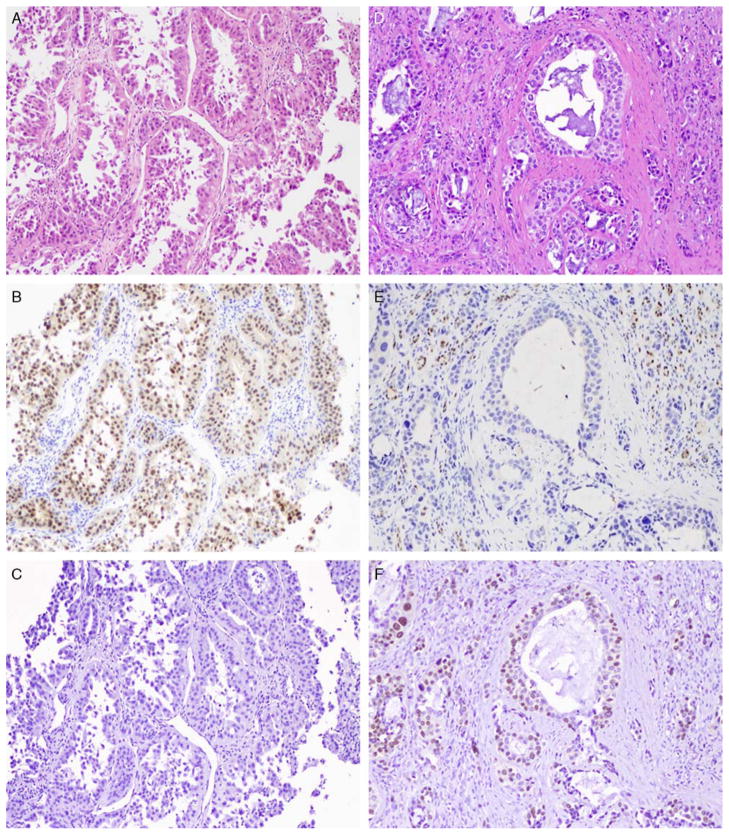
Background: Collecting duct carcinoma (CDC) is a relatively rare but aggressive type of renal malignancy with variable morphologic features. One of the World Health Organization diagnostic criteria for CDC is the exclusion of urothelial carcinoma of renal pelvis from the differential diagnosis. PAX8 is a novel lineage restricted transcription factor expressed in renal tubules. We investigated the expression pattern of PAX8 in CDC and its utility, in combination with p63, in resolving the differential diagnosis of CDC versus upper tract urothelial carcinoma (UUC).
Design: Archival tissues from 21 CDC and 34 UUC were retrieved from our institutional files. Immunohistochemistry for PAX8 and p63 were performed on routine and tissue microarray sections using standard immunohistochemistry protocol. Intensity of nuclear staining was evaluated for each marker and assigned an incremental 0, 1+, 2+, and 3+ score. Extent of staining was categorized as focal (<25%), nonfocal (25% to 75%), or diffuse (>75%).
Results: CDC: All 21 (100%) CDC were positive for PAX8. Intensity of expression was moderate to strong (2+/3+) in 19 cases (90%). Extent of staining was diffuse in 13 of 21 tumors. The p63 was positive in 3 of 21 (14%) CDC cases (PAX8+/p63+). UUC: The 34 UUC included 5 pT1, 4 pT2, and 25 pT3/pT4 tumors. Thirty-one of 34 (91.2%) UUC were negative for PAX8, whereas 33 of 34 (97%) were p63 positive. Staining intensity was moderate in 15 cases (44%), of which 12 were nonfocal or diffuse. The unique p63-negative UUC was a pT1 tumor that was also negative for PAX8 (PAX8-/p63-).
Conclusions: We propose the use of the combination of PAX8 and p63 in the diagnosis of poorly differentiated renal sinus epithelial neoplasms where the differential diagnosis includes CDC versus UUC. The immunoprofile of PAX8+/p63- supports the diagnosis of CDC with a sensitivity of 85.7% and a specificity of 100%. In contrast, a (PAX8-/p63+) profile supports the diagnosis of UUC with a sensitivity of 88.2% and a specificity of 100%. The inverse PAX8/p63 expression seen in CDC and UUC supports a renal tubular rather than an urothelial differentiation in CDC given the nephric lineage restriction of PAX8.
Figures
References
-
- Albadine R, Schultz L, Fajardo DA, et al. PAX8 expression in urothelial neoplasia—an immunohistochemical study of 236 cases. Mod Pathol. 2010;23:174A.
-
- Cuckow PM, Nyirady P, Winyard PJ. Normal and abnormal development of the urogenital tract. Prenat Diagn. 2001;21:908–916. - PubMed
-
- Eble JN, Sauter G, Epstein JI, et al. Pathology and genetics of tumours of the urinary system and male genital organs. In: Srigley JR, Moch H, editors. WHO Classification of Tumours. Vol. 2004. IARC Press; Lyon: 2004. pp. 33–34.
-
- Fedor HL, De Marzo AM. Practical methods for tissue microarray construction. Methods Mol Med. 2005;103:89–101. - PubMed
-
- Gupta R, Paner GP, Amin MB. Neoplasms of the upper urinary tract: a review with focus on urothelial carcinoma of the pelvicalyceal system and aspects related to its diagnosis and reporting. Adv Anat Pathol. 2008;15:127–139. - PubMed
MeSH terms
Substances
Grants and funding
LinkOut - more resources
Full Text Sources
Other Literature Sources
Medical
