Regenerate healing outcomes in unilateral mandibular distraction osteogenesis using quantitative histomorphometry
- PMID: 20463629
- PMCID: PMC4608224
- DOI: 10.1097/PRS.0b013e3181e3b351
Regenerate healing outcomes in unilateral mandibular distraction osteogenesis using quantitative histomorphometry
Abstract
Background: The authors' goal was to ascertain regenerate bone-healing metrics using quantitative histomorphometry at a single consolidation period.
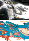
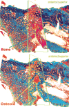
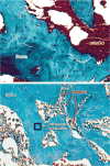
Methods: Rats underwent either mandibular distraction osteogenesis (n = 7) or partially reduced fractures (n = 7); their contralateral mandibles were used as controls (n = 11). External fixators were secured and unilateral osteotomies performed, followed by either mandibular distraction osteogenesis (4 days' latency, then 0.3 mm every 12 hours for 8 days; 5.1 mm) or partially reduced fractures (fixed immediately postoperatively; 2.1 mm); both groups underwent 4 weeks of consolidation. After tissue processing, bone volume/tissue volume ratio, osteoid volume/tissue volume ratio, and osteocyte count per high-power field were analyzed by means of quantitative histomorphometry.
Results: Contralateral mandibles had statistically greater bone volume/tissue volume ratio and osteocyte count per high-power field compared with both mandibular distraction osteogenesis and partially reduced fractures by almost 50 percent, whereas osteoid volume/tissue volume ratio was statistically greater in both mandibular distraction osteogenesis specimens and partially reduced fractures compared with contralateral mandibles. No statistical difference in bone volume/tissue volume ratio, osteoid volume/tissue volume ratio, or osteocyte count per high-power field was found between mandibular distraction osteogenesis specimens and partially reduced fractures.
Conclusions: The authors' findings demonstrate significantly decreased bone quantity and maturity in mandibular distraction osteogenesis specimens and partially reduced fractures compared with contralateral mandibles using the clinically analogous protocols. If these results are extrapolated clinically, treatment strategies may require modification to ensure reliable, predictable, and improved outcomes.
Conflict of interest statement
Figures


Similar articles
-
Quantitative histologic evidence of amifostine-induced cytoprotection in an irradiated murine model of mandibular distraction osteogenesis.Plast Reconstr Surg. 2012 Dec;130(6):1199-1207. doi: 10.1097/PRS.0b013e31826d2201. Plast Reconstr Surg. 2012. PMID: 22878481 Free PMC article.
-
Analysis of the biomechanical properties of the mandible after unilateral distraction osteogenesis.Plast Reconstr Surg. 2010 Aug;126(2):533-542. doi: 10.1097/PRS.0b013e3181de2240. Plast Reconstr Surg. 2010. PMID: 20375764 Free PMC article.
-
Biomechanical assessment of regenerate integrity in irradiated mandibular distraction osteogenesis.Plast Reconstr Surg. 2009 Feb;123(2 Suppl):114S-122S. doi: 10.1097/PRS.0b013e318191c5d2. Plast Reconstr Surg. 2009. PMID: 19182670
-
H Vessel Formation as a Marker for Enhanced Bone Healing in Irradiated Distraction Osteogenesis.Semin Plast Surg. 2024 Jan 19;38(1):31-38. doi: 10.1055/s-0043-1778039. eCollection 2024 Feb. Semin Plast Surg. 2024. PMID: 38495069 Free PMC article. Review.
-
Overview of biological mechanisms and applications of three murine models of bone repair: closed fracture with intramedullary fixation, distraction osteogenesis, and marrow ablation by reaming.Curr Protoc Mouse Biol. 2015 Mar 2;5(1):21-34. doi: 10.1002/9780470942390.mo140166. Curr Protoc Mouse Biol. 2015. PMID: 25727198 Free PMC article. Review.
Cited by
-
Dose-response effect of human equivalent radiation in the murine mandible: part I. A histomorphometric assessment.Plast Reconstr Surg. 2011 Jul;128(1):114-121. doi: 10.1097/PRS.0b013e31821741d4. Plast Reconstr Surg. 2011. PMID: 21701328 Free PMC article.
-
Quantitative histologic evidence of amifostine-induced cytoprotection in an irradiated murine model of mandibular distraction osteogenesis.Plast Reconstr Surg. 2012 Dec;130(6):1199-1207. doi: 10.1097/PRS.0b013e31826d2201. Plast Reconstr Surg. 2012. PMID: 22878481 Free PMC article.
-
Dose-response effect of human equivalent radiation in the murine mandible: Part II. A biomechanical assessment.Plast Reconstr Surg. 2011 Nov;128(5):480e-487e. doi: 10.1097/PRS.0b013e31822b67ae. Plast Reconstr Surg. 2011. PMID: 22030507 Free PMC article.
-
A Histomorphometric Analysis of Radiation Damage in an Isogenic Murine Model of Distraction Osteogenesis.J Oral Maxillofac Surg. 2015 Dec;73(12):2419-28. doi: 10.1016/j.joms.2015.08.002. Epub 2015 Aug 7. J Oral Maxillofac Surg. 2015. PMID: 26341682 Free PMC article.
-
Amifostine remediates the degenerative effects of radiation on the mineralization capacity of the murine mandible.Plast Reconstr Surg. 2012 Apr;129(4):646e-655e. doi: 10.1097/PRS.0b013e3182454352. Plast Reconstr Surg. 2012. PMID: 22456378 Free PMC article.
References
-
- Reddy LV, Elhadi HM. Maxillary advancement by distraction osteogenesis. Atlas Oral Maxillofac Surg Clin North Am. 2008;16:237–247. - PubMed
-
- Nishimoto S, Oyama T, Nagashima T, et al. Lateral orbital expansion and gradual fronto-orbital advancement: An option to treat severe syndromic craniosynostosis. J Craniofac Surg. 2008;19:1622–1627. - PubMed
-
- Wong GB, Ciminello FS, Padwa BL. Distraction osteogenesis of the cleft maxilla. Facial Plast Surg. 2008;24:467–471. - PubMed
-
- Yu JC, Fearon J, Havlik RJ, Buchman R, Polley JW. Distraction osteogenesis of the craniofacial skeleton. Plast Reconstr Surg. 2004;114:1E–20E. - PubMed
-
- Kamoshima Y, Sawamura Y, Yoshino M, Kawashima K. Frontofacial monobloc advancement using gradual bone distraction method. J Pediatr Surg. 2008;43:1944–1948. - PubMed
Publication types
MeSH terms
Grants and funding
LinkOut - more resources
Full Text Sources
Medical

