. 2010 Dec;78(11):1088-99.
doi: 10.1038/ki.2010.136. Epub 2010 May 12.

Podocytes require the engagement of cell surface heparan sulfate proteoglycans for adhesion to extracellular matrices

Affiliations

Podocytes require the engagement of cell surface heparan sulfate proteoglycans for adhesion to extracellular matrices

Shoujun Chen et al. Kidney Int. 2010 Dec.

Abstract

Podocytes adhere to the glomerular basement membrane by cell surface receptors. Since in other cells these adhesions are enhanced by cell surface proteoglycans, we examined the contribution of these molecules and their glycosaminoglycan side chains to podocyte adhesion by developing immortalized podocyte cell lines with (control) or without (mutant) heparan sulfate glycosaminoglycan chains. In adhesion assays control podocytes attached, spread, and migrated more efficiently compared with mutants, indicating a requirement for heparan sulfate chains in these processes. The proteoglycan syndecan-4 is known to have direct effects on cell attachment, spreading, and cytoskeletal organization. We found it localized to focal adhesions in control podocytes coincident with stress fiber formation. In mutant cells, syndecan-4 was associated with smaller focal contacts and cortical actin organization. Analysis by flow cytometry showed that mutant cells had twice the amount of surface syndecan-4 of control cells. Protein kinase Cα, a signaling molecule bound to and activated by syndecan-4, showed a fourfold increase in membrane localization-activation than that seen in control cells. In vivo, the loss of heparan sulfate glycosaminoglycans in PEXTKO mice led to a loss of glomerular syndecan-4. Overall, our study provides further evidence for a dynamic role of cell surface heparan sulfate glycosaminoglycans in podocyte activity.


Figures
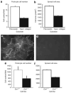
Figure 1
Figure 1. Podocytes use cell-surface heparan sulfate proteoglycans (HSPGs) for cell–matrix interactions
To initially analyze the possibility that the cell surface HS may have a role in podocyte attachment, adhesion assays using EXT1+/+ podocytes were run using substrata consisting of either monomeric type I collagen or plasma fibronectin. The data show that podocytes attach (a) and spread (b) (P<0.0001) less efficiently on monomeric type I collagen compared with fibronectin. The micrographs shown in (c) and (d) are of immortalized EXT1fl/fl podocytes (c) that were infected by adenoviral-mediated gene transfer with constructs of Ad-green fluorescent protein (GFP) (HS+ podocytes) or (d) GFP-Cre recombinase (HS− podocytes). The images show the cells from cultures of both lines 48 h after seeding. Panel (c) shows HS4C3 staining (anti-HS) of the HS rich extracellular matrix laid down by podocytes during the timeframe. In the Cre-recombinase-transfected cells (d), there is no staining by HS4C3 at either the cell surface or the pericellular matrix. (Final magnification × 200, the exposure time for both panels was held constant.) The graphs in Figure 2e and f shows that the loss of cell surface HS (EXT1−/−) in podocytes results in a significant decrease in cell adhesion (e, P<0.01) and spread cell area (f, P<0.0001) compared with control cells (EXT1+/+).
Figure 2
Figure 2. Loss of cell surface heparan sulfate (HS) delays podocyte cell migration in a scratch wound assay system
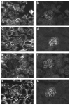
Confluent monolayers of HS+ (a) or HS− (b) cells grown in 24-well plates were scratch-wounded in a crosswise manner using a pipette tip. Registration marks (R in micrographs) were etched into the bottom side of the plate to facilitate later image alignments during the course of the study. The progress of cell migration into the denuded area tracked over time using phase contrast microscopy. Images were taken of the boundaries of the wound areas at T = 0 (red line), T = 24 (blue line), and T = 48 (green line) and the leading edges of the cell migration traced using image analysis software (final magnification × 100). The area for each scratch wound was calculated using a planimetry subroutine in the image analysis software and the relative change in area from T = 0 was determined using the equation [A0An]A0=ΔA, where A0 = area at T=0, An = area at Time = n, and ΔA is the relative change in area. (c) is the bar graph depicting the relative change in the wound area as a function of time. At both times (T = 24, 48) the HS+ cells showed a greater efficiency (0.32 for HS+ vs 0.24 for HS− at T = 24; 0.65 for HS+ vs 0.53 for HS− at T = 48) for wound closure (P<0.006 for T = 24 h, P<0.03 T = 48 h).
Figure 3
Figure 3. Loss of heparan sulfate (HS) in podocytes affects cytoskeletal organization and focal adhesion assembly in podocytes in short-term adhesion assays
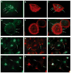
Podocytes were seeded at low density (10K cells per well) on a fibronectin substratum for 4 h). The cells were double-label immunostained with antibodies directed against syndecan-4 (a, d, g, j, green) or vinculin (h, k, red) or the actin cytoskeleton stained with fluorchrome-conjugated phalloidin (b, e, red). Hoechst 33242 nuclear stain was used as a nuclear counterstain in some cultures. (c, f, i, l) are the digital overlay images for each respective row. HS+ podocytes stained showed prominent stress fibers (b), some of which appeared to terminate in large, discrete clusters of syndecan-4 (a, arrows). Cortical actin was observed (e) in HS− podocytes that were able to spread onto the fibronectin substrate. Syndecan-4 staining in these cells (d) appeared as small punctate but more numerous clusters around the periphery of the cell (arrows). In HS+ cells that were actively spreading (g–i), syndecan-4 staining (g) colocalized with vinculin staining (h) in cell processes (arrows); some actively spreading podocytes that were HS− (j–l) had multiple thin processes extending from the cell body, the termini of which showed colocalization for vinculin (k) and syndecan-4 (j, arrows). (Final magnification × 400.)
Figure 4
Figure 4. The pattern of localization of known syndecan-4-binding proteins is altered in heparan sulfate (HS)-GAG− podocytes
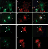
The lamellipodia of actively spreading HS+ podocytes showed large, discrete clustering of syndecan-4 (a, arrowheads), some of which colocalized with α actinin-4 staining (b, arrowheads). Staining for α actinin-4 also appeared as fibrils radiating from the areas of syndecan-4-positive lamellipodia (b, smaller arrows) toward the center of the cell. In cells that appeared to be in the earlier stages of lamellipodia extension both syndecan-4 and -actinin-4 were colocalized at the leading edge (a, b, larger arrows). In HS− podocytes (d), syndecan-4 stained the ends of small processes (arrows) and colocalized with α actinin-4 (e, arrows) in these processes. Protein kinase Cα (h) staining was observed at the leading edges of lamellipodia (arrowheads), colocalized with syndecan-4 (g, arrowheads) in HS+ podocytes (gi). In HS− podocytes (jl) both PKCα (k) and syndecan-4 (j) were colocalized (arrowhead) in small membrane ruffles and small processes extending from the cell body. (Final magnification × 400.)
Figure 5
Figure 5. The expression of syndecan-4 is upregulated in heparan sulfate (HS)− podocytes when compared with HS + podocytes
(a and b) show HS+ (a) and HS− (b) podocytes immunostained syndecan-4 in the process of adhesion to fibronectin-coated (100 μg/ml) substratum at T=2 h. The HS+ podocytes have attached and spread, the arrows (white) indicating focal clusters of syndecan-4 immunoreactivity at the leading edges of the cells. The HS− podocytes during the same time interval have attached but are poorly spread, the arrows indicating small processes extending from the round cell body. (Final magnification × 200.) The graph and chart in c represent the results of flow cytometry studies. The graph shows three traces: non-immune antibody control formula image, HS + cells stained for syndecan-4 formula image, and HS− cells stained for syndecan-4 formula image. The chart shows the relative fluorescence intensity values for syndecan-4 staining in HS+ and HS− cells. The data show a net increase in the staining for syndecan-4 in HS− cells.
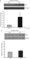
Figure 6
Figure 6. The membrane localization of PKCα is significantly enhanced in heparan sulfate (HS)− podocytes when compared with HS+ podocytes
Isolated membrane (a) and cytosol (b) fractions from HS+ and HS− podocytes were separated by sodium dodecyl sulfate-polyacrylamide gel electrophoresis (SDS-PAGE) and the respective pools analyzed by western blot immunoassay using antibodies recognizing PKCα. The blot was subsequently reprobed with antibodies against β1 integrin (for cell membranes) or glyceraldehyde 3-phosphate dehydrogenase (for cytosol) as a normalization control. The graph below shows the results of densitometric scanning of the blot, the PKCα membrane localization/activation significantly greater (P<0.001) in the HS− cells compared with the HS+ cells. In contrast, there were no significant differences between the two cell populations with regard to cytosolic levels of PKCα.
Figure 7
Figure 7. Loss of heparan sulfate (HS)-GAGs in podocytes alters the distribution of syndecan-4 and PKCα in confluent monolayers of podocytes
Podocytes were seeded at high density (25K cells per well) on a fibronectin substratum. After fixation and permeablilization the cells were immunostained with antibodies directed against syndecan-4 (b, e) and PKCα (a, d). (c, f) represent digital overlays of the images in their respective rows. The images show that in the HS+ cells syndecan-4 and PKCα colocalize in a linear pattern at the edges of cells in which cell–cell contact is made (ac). In HS− podocytes (df), the peripheral localization of PKCα and syndecan-4 was entirely disrupted, only focal clusters of colocalization are observed. (Final magnification × 400.)
Figure 8
Figure 8. Podocytes make syndecan-4 in vivo
The micrographs in Figure 8 are sections of kidney immunostained for syndecan-4 (a, c, e, g) and for synaptopodin (b, d, f, h). (a, b) are glomeruli from controls (heparan sulfate (HS) +) 2.5PCre-Ext1+/+ animals; (c, d) are glomeruli from controls (agrin +) 2.5PCre-Agrn+/fl animals. In both control groups, syndecan-4 (a, c) immunostaining within the glomerulus is seen as a punctate pattern that colocalizes (arrows) in part with synaptopodin (b, d). The same pattern of localization is also maintained in the 2.5PCre-Agrnfl/fl mice (g), whose podocytes are unable to make the full-length core protein of agrin. In contrast, the punctate pattern of syndecan-4 immunostaining is severely diminished in the glomeruli from the 2.5PCre-Ext1fl/fl mice (e) (final magnification × 400).

Similar articles

Cited by

References

    1. Kanwar YS, Farquhar MG. Anionic sites in the glomerular basement membrane. In vivo and in vitro localization to the lamina rarae by cationic probes. J Cell Biol. 1979;81:137–153. - PMC - PubMed
    1. Kanwar YS, Linker A, Farquhar MG. Increased permeability of the glomerular basement membrane to ferritin after removal of glycosaminoglycans (heparan sulfate) by enzyme digestion. J Cell Biol. 1980;86:688–693. - PMC - PubMed
    1. Harvey SV, Jarad G, Cunningham J, et al. Disruption of glomerular basement membrane charge through podocyte-specific mutation of agrin does not alter glomerular permselectivity. Am J Path. 2007;171:139–152. - PMC - PubMed
    1. Chen S, Wassenhove-McCarthy DJ, Yamaguchi Y, et al. Loss of heparan sulfate glycosaminoglycan assembly in podocytes does not lead to proteinuria. Kidney Int. 2008;74:289–299. - PMC - PubMed
    1. Woods A. Syndecans: transmembrane modulators of adhesion and matrix assembly. J Clin Invest. 2001;107:935–941. - PMC - PubMed

Publication types

MeSH terms

LinkOut - more resources