. 2010 May 6;6(5):e1000877.
doi: 10.1371/journal.ppat.1000877.

Prolonged antigen presentation is required for optimal CD8+ T cell responses against malaria liver stage parasites

Affiliations

Prolonged antigen presentation is required for optimal CD8+ T cell responses against malaria liver stage parasites

Ian A Cockburn et al. PLoS Pathog. .

Abstract

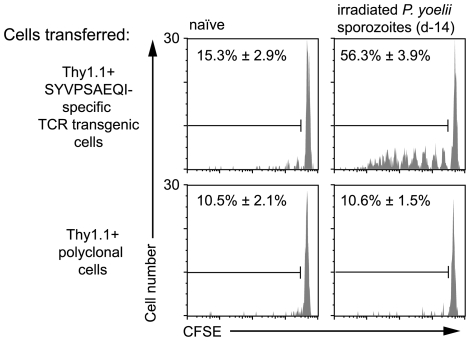
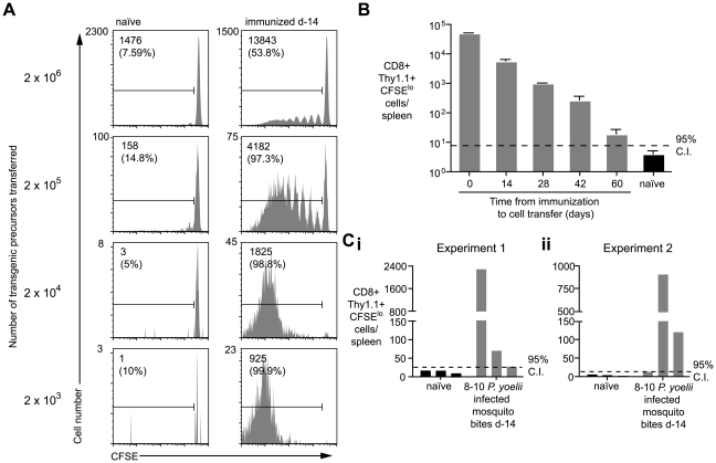
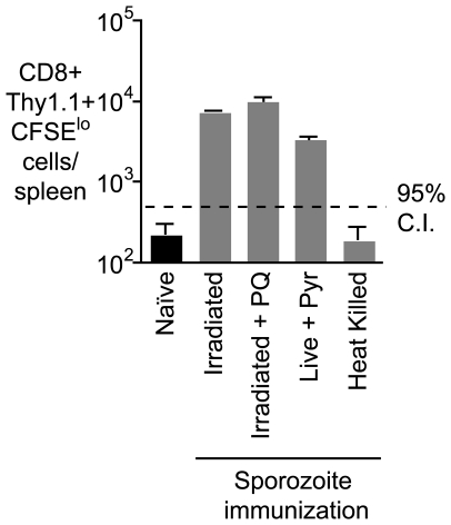
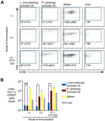
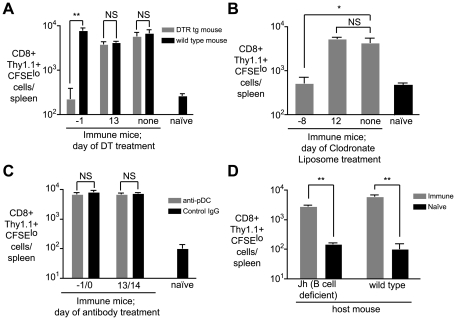
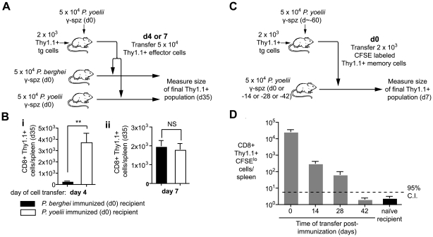
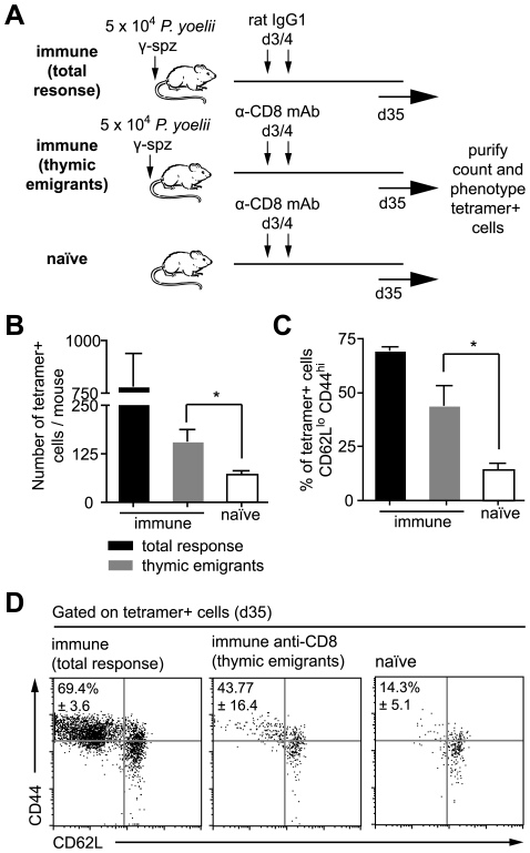
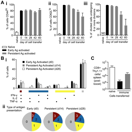
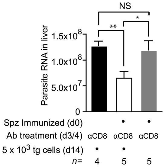
Immunization with irradiated sporozoites is currently the most effective vaccination strategy against liver stages of malaria parasites, yet the mechanisms underpinning the success of this approach are unknown. Here we show that the complete development of protective CD8+ T cell responses requires prolonged antigen presentation. Using TCR transgenic cells specific for the malaria circumsporozoite protein, a leading vaccine candidate, we found that sporozoite antigen persists for over 8 weeks after immunization--a remarkable finding since irradiated sporozoites are incapable of replication and do not differentiate beyond early liver stages. Persisting antigen was detected in lymphoid organs and depends on the presence of CD11c+ cells. Prolonged antigen presentation enhanced the magnitude of the CD8+ T cell response in a number of ways. Firstly, reducing the time primed CD8+ T cells were exposed to antigen in vivo severely reduced the final size of the developing memory population. Secondly, fully developed memory cells expanded in previously immunized mice but not when transferred to naïve animals. Finally, persisting antigen was able to prime naïve cells, including recent thymic emigrants, to become functional effector cells capable of eliminating parasites in the liver. Together these data show that the optimal development of protective CD8+ T cell immunity against malaria liver stages is dependent upon the prolonged presentation of sporozoite-derived antigen.


Conflict of interest statement

The authors have declared that no competing interests exist.

Figures

Figure 1
Figure 1. Prolonged antigen presentation after sporozoite immunization.
2×10⁶ Thy1.1+ CD8+ T cells from either TCR transgenic or WT animals were transferred into groups of naïve mice or mice immunized 2 weeks previously with 5×10⁴ irradiated P. yoelii sporozoites. 10 days after cell transfer the spleen cells from the recipient mice were analyzed by FACs. Histograms show CFSE profiles of the Thy1.1+ CD8+ cell populations in each group. Values are the mean ± SE of the percentage of divided cells (n = 3, data from one of two similar experiments shown).
Figure 2
Figure 2. Duration of antigen presentation, and antigen persistence after mosquito biting.
A. Mice were immunized with 5×10⁴ irradiated sporozoites i.v. and 14 days later 2×10³–2×10⁶ CFSE labeled transgenic cells were transferred to the mice and to unimmunized controls. Ten days later transgenic cells were enriched from the spleen and their CFSE profile determined by FACs. Representative plots from one of three mice per group are shown. Values given are the total number of divided transgenic (CD8+ Thy1.1+ CFSElo) cells isolated from the spleen and in parentheses the % of total recovered transgenic cells that have divided (data representative of numerous similar experiments). B. Mice were immunized as in A. and 1×10⁴ transgenic cells were transferred to mice at different time points after immunization. Ten days after transfer the cells were recovered and the number of divided transgenic cells recovered was determined (n = 3, mean ± SE; data representative of two similar experiments). C. Three mice were bitten by 8–10 infected P. yoelii infected mosquitoes. Fourteen days later 1×10⁴ transgenic cells were transferred to the mice and 10 days later the number of divided transgenic cells was determined. Bars represent values for individual mice; data shown from two independent experiments (i and ii).
Figure 3
Figure 3. Prolonged antigen presentation does not depend on the presence of parasite.
Mice were immunized i.v. with 5×10⁴ of each of irradiated sporozoites, irradiated sporozoites followed by 2 doses of PQ treatment 10 hours apart 7 days after immunization, live sporozoites (with pyrimethamine treatment to remove blood stage parasites) and heat killed sporozoites,. Fourteen days later 2×10⁵ transgenic cells were transferred to the immunized and naïve mice, 10 days after transfer the number of transgenic cells/ spleen was determined for each group of mice (n = 3, mean ± SE; data from one of three experiments).
Figure 4
Figure 4. Antigen persists in the draining lymph nodes.
A. Mice were immunized i.v. or i.d. with 5×10⁴ irradiated sporozoites. Fourteen days later 1×10⁴ CFSE labeled transgenic cells were transferred to the mice, some of the mice that had been immunized i.d. received daily doses of 30 µg FTY720 after cell transfer to prevent egress of primed T cells. Eight days after cell transfer transgenic cells were enriched from various tissues by positive selection. Flow cytometry plots show representative data from each group. Values are the mean ± standard deviation of the number of divided transgenic cells recovered per tissue of three mice per group. B. Histograms of the data presented in A. (n = 3, mean ± SE, P>0.05, * = P<0.05, *** = P<0.001). Data in A and B are representative of four similar experiments.
Figure 5
Figure 5. CD11c+ and phagocytic cells are required for continued antigen presentation.
A. CD11c-DTR and WT mice were immunized i.v. with 5×10⁴ irradiated sporozoites at day 0. Groups of CD11c-DTR and wild-type mice were treated with 100ng of DT on the indicated days. On day 14, 2×10⁵ CFSE labeled transgenic cells were transferred to the mice. Ten days later, the number of divided transgenic cells was determined by FACs (n = 3, mean ± SE, * = P<0.05, ** = P<0.01, data representative of four experiments). B. Groups of mice were immunized on day 0 as in A and treated with clodronate liposomes (200 µl i.v./mouse) at the indicated days to deplete splenic macrophage populations. On day 14, 2×10⁵ CFSE labeled transgenic cells were transferred to the mice. Ten days later, the number of divided transgenic cells was determined by FACs (n = 3, mean ± SE, * = P<0.05 data representative of two experiments). C. Groups of mice were immunized on day 0 with irradiated sporozoites and treated with 200 µg of the anti-plasmacytoid DC mAb 120G8 or control IgG at the indicated days. On day 14, 2×10⁵ transgenic cells were transferred to the mice. Ten days later the number of divided transgenic cells was determined by FACs. (n = 3, mean ± SE, data representative of two experiments). D. Jh (B cell deficient) and WT mice were immunized with irradiated sporozoites. 14 days later 2×10⁵ transgenic cells were transferred to the mice. Ten days after cell transfer the number of divided transgenic cells in immune mice was determined by FACs and compared to background proliferation of cells transferred to naïve mice (n = 3, mean ± SE, ** = P<0.01, data representative of two experiments).
Figure 6
Figure 6. Prolonged antigen presentation is required for optimal CD8+ memory formation.
A and B. Naïve mice received 2×10³ transgenic cells and were immunized on d0 with 5×10⁴ irradiated P. yoelii sporozoites. On either d4 or d7 CD8+ T cells were purified from these mice and the equivalent of 5×10⁴ transgenic cells were transferred to recipients that had received 5×10⁴ irradiated P. yoelii sporozoites or 5×10⁴ irradiated P. berghei sporozoites on d0. On d35 the size of the transgenic memory population was measured by FACs. A. Outline of the experiment. B. Size of the resulting memory populations after cells transfer on (i) d4, (ii) d7 (n = 5, mean ± SE, ** = P<0.01). C and D. BALB/c mice were immunized with 5×10⁴ irradiated sporozoites and 2×10³ d60 memory transgenic cells were transferred to mice at different time points after immunization. Ten days after transfer the transgenic cells were recovered and the number of divided cells was determined C. Outline of the experiment. D Number of divided cells recovered (n = 3, mean ± SE; a positive response is considered > upper 95% confidence interval of the response seen in naïve mice, data from one of two similar experiments).
Figure 7
Figure 7. Persisting antigen primes naïve thymic emigrants.
A. Outline of the experiment: mice were immunized with 5×10⁴ irradiated sporozoites i.v. On days 3 and 4 after immunization some of the mice were depleted of CD8+ cells such that the observed response consists largely of later thymic emigrants (gray bars). Control mice were treated with control IgG1 leaving the pre-existing response in tact (total response – black bars), while other mice were left un-immunized but treated with anti-CD8 (naïve – white bars). B. Histograms of the number of tetramer+ cells recovered in each group, bar charts show means ± SE (n = 6, * = P<0.05, data pooled from two similar experiments). C. Histograms of the percentage of activated cells (CD62Llo, CD44hi) among the tetramer+ population recovered in each experiment (n = 3, * = P<0.05, data from one of two similar experiments). D. Dot plots of the CD62L and CD44 expression of tetramer+ cells in each group, data show concatenated data from 3 animals per group and are from one of two similar experiments.
Figure 8
Figure 8. Antigen presentation weeks after immunization can prime T cells to become functional effector cells.
A. 2×10³ CFSE labeled transgenic cells were transferred to mice that had been immunized 14, 28, 42 and 60d previously with 5×10⁴ irradiated sporozoites i.v. (persistent-antigen activated cells – gray bars). Phenotypes of persistent antigen activated cells were compared to the phenotypes of early antigen activated cells, in which 2×10³ transgenic cells were transferred to naïve mice and immunized the same day with 5×10⁴ irradiated sporozoites (black bars). Naïve control mice received 2×10⁶ CFSE labeled transgenic cells. 7 days after cell transfer transgenic cells were isolated and analyzed for (i) CD44 expression, (ii) CD62L expression, and (iii) CFSE dilution (n = 3, means ± SE, ns = not significant, * = P<0.05, ** P<0.01, data from four similar experiments). B. Polyfunctional analysis comparing the cytokine expression of cells stimulated by early (d0) or persisting antigen (d14 or d28) 35 days after cell transfer. i. histogram shows the % of transgenic cells producing each possible combination of cytokines in response to early antigen presentation (black bars), d14 antigen presentation (gray bars) or d28 antigen presentation (white bars) ii. pie charts representing the number of cells producing 3 (red), 2 (blue), 1 (yellow) or 0 (gray) cytokines (data from three similar experiments). C. 2×10³ naïve transgenic cells (white bar), 2×10³ d35 memory transgenic cells primed by early antigen (black bar) and 2×10³ d35 memory transgenic cells primed by persisting antigen (gray bar) were transferred to naïve BALB/C mice which were immunized with 2×10⁶ VV-SYV i.v., 14 days later the number of Thy1.1+ transgenic cells was measured by FACs (n = 3, mean ± SE, data from two similar experiments).
Figure 9
Figure 9. Cells primed weeks after immunization can inhibit parasite development.
Two groups of mice were immunized with 3×10⁴ irradiated sporozoites i.d. in the right ear on d0 and depleted of the endogenous CD8+ T cell response on days 3 and 4. Some of these mice received 2×10³ transgenic cells on d14 (white bar) while others did not receive cells (gray bar). Naïve control mice were similarly depleted of CD8+ T cells on days 3 and 4 and received transgenic cells (black bar). All mice were then challenged on d21 and euthanized 40 hours later. Parasite burdens in the liver were measured by RT-PCR (mean ± SE, * = P<0.05, ** = P<0.01, data from one of two experiments shown).

Similar articles

Cited by

References

    1. Clyde DF. Immunization of man against falciparum and vivax malaria by use of attenuated sporozoites. Am J Trop Med Hyg. 1975;24:397–401. - PubMed
    1. Nussenzweig RS, Vanderberg J, Most H, Orton C. Protective immunity produced by the injection of x-irradiated sporozoites of plasmodium berghei. Nature. 1967;216:160–162. - PubMed
    1. Schofield L, Villaquiran J, Ferreira A, Schellekens H, Nussenzweig R, et al. Gamma interferon, CD8+ T cells and antibodies required for immunity to malaria sporozoites. Nature. 1987;330:664–666. - PubMed
    1. Weiss WR, Sedegah M, Beaudoin RL, Miller LH, Good MF. CD8+ T cells (cytotoxic/suppressors) are required for protection in mice immunized with malaria sporozoites. Proc Natl Acad Sci U S A. 1988;85:573–576. - PMC - PubMed
    1. Scheller LF, Azad AF. Maintenance of protective immunity against malaria by persistent hepatic parasites derived from irradiated sporozoites. Proc Natl Acad Sci U S A. 1995;92:4066–4068. - PMC - PubMed

Publication types

MeSH terms

Substances