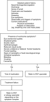
Diagnostic strategies in nasal congestion
- PMID: 20463824
- PMCID: PMC2866556
- DOI: 10.2147/ijgm.s8084
Diagnostic strategies in nasal congestion
Abstract
Nasal congestion is a major symptom of upper respiratory tract disorders, and its characterization an important part of the diagnosis of these illnesses. Patient history and assessment of nasal symptoms are essential components of diagnosis, providing an initial evaluation that may be adequate to rule out serious conditions. However, current congestion medications are not always fully effective. Thus, if symptoms do not respond adequately to therapy, or symptoms suggestive of more serious conditions are present, specialized assessments may be needed. Various techniques are available for diagnosing patients, including those used chiefly by primary care clinicians and those requiring the expertise of otolaryngologists, allergists, and other specialists. Endoscopy remains a mainstay for evaluating nasal blockage and its causes, while modalities such as peak nasal inspiratory flow and acoustic rhinometry are evolving to provide easy-to-use, noninvasive procedures that are sensitive enough to measure small but clinically important abnormalities and therapeutic changes. Several imaging modalities are available to the specialist for severe or unusual cases, as are specialized diagnostic procedures that measure adjunctive features of congestion, such as impaired mucociliary function.
Keywords: allergic rhinitis; congestion; diagnosis; obstruction; rhinosinusitis.
Figures


Similar articles
-
Objective measurement of nasal airway dimensions using acoustic rhinometry: methodological and clinical aspects.Allergy. 2002;57 Suppl 70:5-39. doi: 10.1046/j.0908-665x.2001.all.doc.x. Allergy. 2002. PMID: 11990714
-
EPOS Primary Care Guidelines: European Position Paper on the Primary Care Diagnosis and Management of Rhinosinusitis and Nasal Polyps 2007 - a summary.Prim Care Respir J. 2008 Jun;17(2):79-89. doi: 10.3132/pcrj.2008.00029. Prim Care Respir J. 2008. PMID: 18438594 Free PMC article. Review.
-
Clinical practice guideline: Allergic rhinitis.Otolaryngol Head Neck Surg. 2015 Feb;152(1 Suppl):S1-43. doi: 10.1177/0194599814561600. Otolaryngol Head Neck Surg. 2015. PMID: 25644617
-
Objective monitoring of nasal patency and nasal physiology in rhinitis.J Allergy Clin Immunol. 2005 Mar;115(3 Suppl 1):S442-59. doi: 10.1016/j.jaci.2004.12.015. J Allergy Clin Immunol. 2005. PMID: 15746882 Free PMC article. Review.
-
Nasal congestion index: A measure for nasal obstruction.Laryngoscope. 2009 Aug;119(8):1628-32. doi: 10.1002/lary.20505. Laryngoscope. 2009. PMID: 19507219
Cited by
-
Allergic Rhinitis in Children: A Randomized Clinical Trial Targeted at Symptoms.Indian J Otolaryngol Head Neck Surg. 2014 Dec;66(4):386-93. doi: 10.1007/s12070-014-0708-4. Epub 2014 Feb 11. Indian J Otolaryngol Head Neck Surg. 2014. PMID: 26396949 Free PMC article.
-
A Comparative Health Utility Value Analysis of Outcomes for Patients Following Septorhinoplasty With Previous Nasal Surgery.JAMA Facial Plast Surg. 2019 Sep 1;21(5):402-406. doi: 10.1001/jamafacial.2019.0176. JAMA Facial Plast Surg. 2019. PMID: 31194223 Free PMC article.
-
Objective assessment of persistent rhinitis in Chinese and its relationship with serum indicators.Eur Arch Otorhinolaryngol. 2015 Jul;272(7):1679-85. doi: 10.1007/s00405-014-3241-x. Epub 2014 Aug 20. Eur Arch Otorhinolaryngol. 2015. PMID: 25135578
-
Correlation Between Objective Evaluation Result of Nasal Congestion and Life Quality in Patients with Acute Rhinosinusitis.Indian J Otolaryngol Head Neck Surg. 2019 Nov;71(Suppl 3):1929-1934. doi: 10.1007/s12070-018-1333-4. Epub 2018 Apr 7. Indian J Otolaryngol Head Neck Surg. 2019. PMID: 31763270 Free PMC article.
-
Accuracy of peak nasal flow to determine nasal obstruction in patients with allergic rhinitis.Acta Otorhinolaryngol Ital. 2022 Apr;42(2):155-161. doi: 10.14639/0392-100X-N1617. Acta Otorhinolaryngol Ital. 2022. PMID: 35612507 Free PMC article.
References
-
- Bousquet J, Van Cauwenberge P, Khaltaev N. Allergic rhinitis and its impact on asthma. J Allergy Clin Immunol. 2001;108(5 Suppl):S147–S334. - PubMed
-
- Fokkens W, Lund V, Mullol J. European position paper on rhinosinusitis and nasal polyps 2007. Rhinol Suppl. 2007;(Suppl 20):1–136. - PubMed
-
- van Spronsen E, Ingels KJ, Jansen AH, Graamans K, Fokkens WJ. Evidence-based recommendations regarding the differential diagnosis and assessment of nasal congestion: using the new GRADE system. Allergy. 2008;63(7):820–833. - PubMed
-
- Baraniuk JN, Kim D. Nasonal reflexes, the nasal cycle, and sneeze. Curr Allergy Asthma Rep. 2007;7:105–111. - PubMed
LinkOut - more resources
Full Text Sources
Other Literature Sources

