

Accelerators, Brakes, and Gears of Actin Dynamics in Dendritic Spines
- PMID: 20463852
- PMCID: PMC2867483
- DOI: 10.2174/1874082000903020067
Accelerators, Brakes, and Gears of Actin Dynamics in Dendritic Spines
Abstract
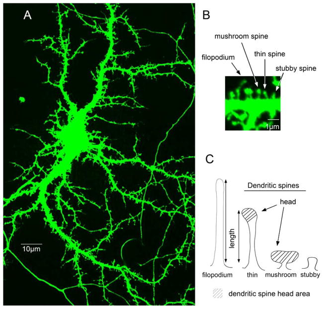
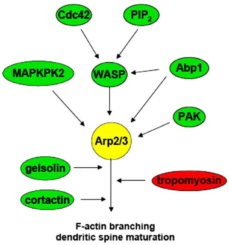
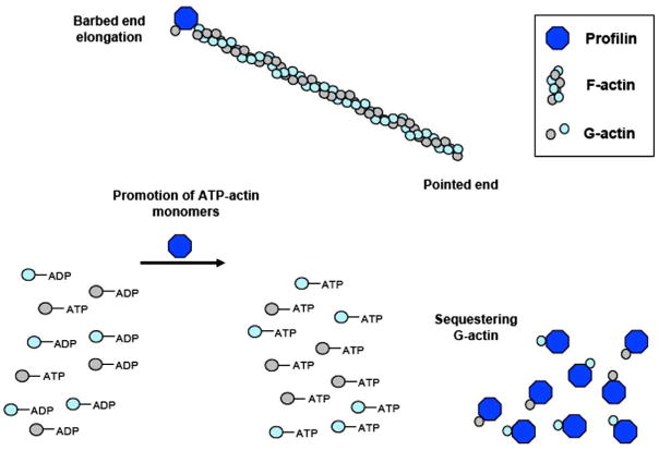
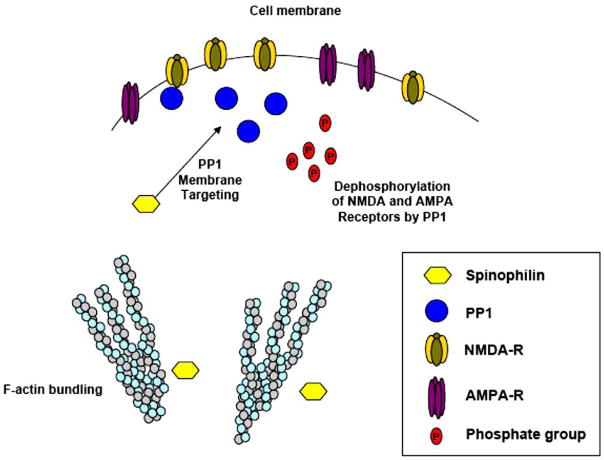
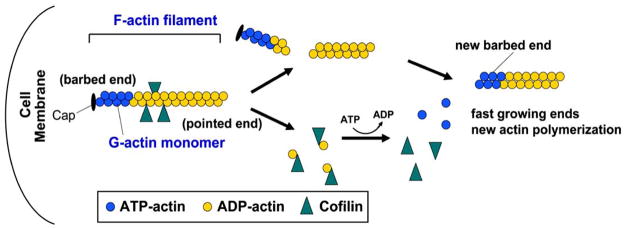
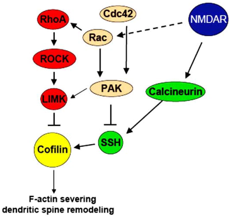
Dendritic spines are actin-rich structures that accommodate the postsynaptic sites of most excitatory synapses in the brain. Although dendritic spines form and mature as synaptic connections develop, they remain plastic even in the adult brain, where they can rapidly grow, change, or collapse in response to normal physiological changes in synaptic activity that underlie learning and memory. Pathological stimuli can adversely affect dendritic spine shape and number, and this is seen in neurodegenerative disorders and some forms of mental retardation and autism as well. Many of the molecular signals that control these changes in dendritic spines act through the regulation of filamentous actin (F-actin), some through direct interaction with actin, and others via downstream effectors. For example, cortactin, cofilin, and gelsolin are actin-binding proteins that directly regulate actin dynamics in dendritic spines. Activities of these proteins are precisely regulated by intracellular signaling events that control their phosphorylation state and localization. In this review, we discuss how actin-regulating proteins maintain the balance between F-actin assembly and disassembly that is needed to stabilize mature dendritic spines, and how changes in their activities may lead to rapid remodeling of dendritic spines.
Figures

References
-
- Rao A, Craig AM. Signaling between the actin cytoskeleton and the postsynaptic density of dendritic spines. Hippocampus. 2000;10:527–41. - PubMed
-
- Sorra KE, Harris KM. Overview on the structure, composition, function, development, and plasticity of hippocampal dendritic spines. Hippocampus. 2000;10:501–11. - PubMed
-
- Hering H, Sheng M. Dendritic spines: structure, dynamics and regulation. Nat Rev Neurosci. 2001;2:880–8. - PubMed
-
- Yuste R, Bonhoeffer T. Genesis of dendritic spines: insights from ultrastructural and imaging studies. Nat Rev Neurosci. 2004;5:24–34. - PubMed
-
- Ethell IM, Pasquale EB. Molecular mechanisms of dendritic spine development and remodeling. Prog Neurobiol. 2005;75:161–205. - PubMed
Grants and funding
LinkOut - more resources
Full Text Sources
Research Materials