. 2010 May 6;6(5):e1000774.
doi: 10.1371/journal.pcbi.1000774.

Spontaneous quaternary and tertiary T-R transitions of human hemoglobin in molecular dynamics simulation

Affiliations

Spontaneous quaternary and tertiary T-R transitions of human hemoglobin in molecular dynamics simulation

Jochen S Hub et al. PLoS Comput Biol. .

Abstract

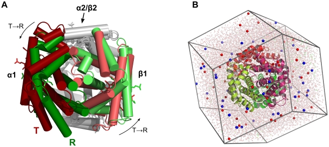
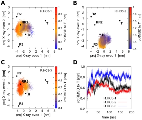
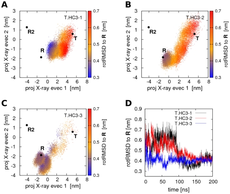
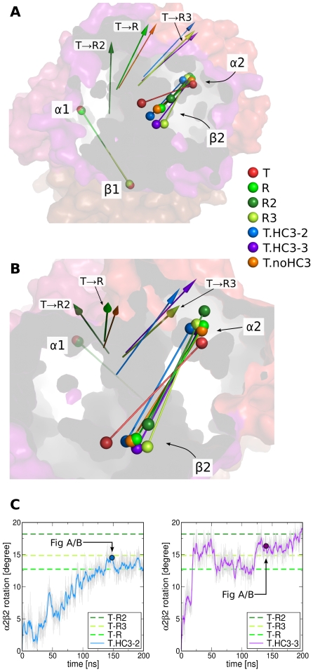
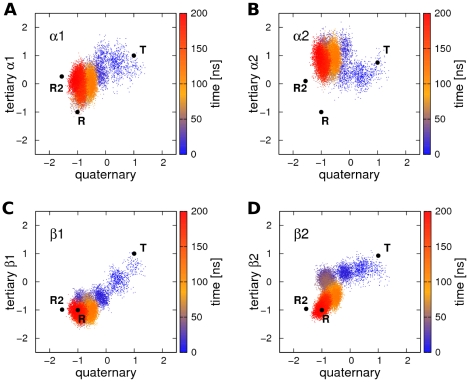
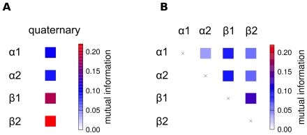
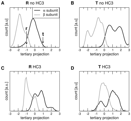
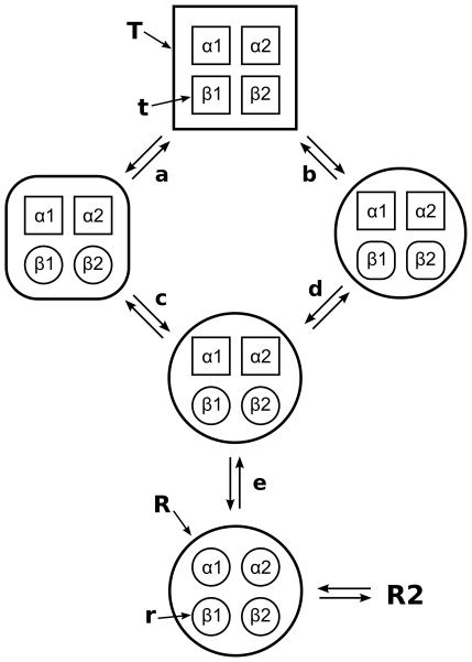
We present molecular dynamics simulations of unliganded human hemoglobin (Hb) A under physiological conditions, starting from the R, R2, and T state. The simulations were carried out with protonated and deprotonated HC3 histidines His(beta)146, and they sum up to a total length of 5.6 micros. We observe spontaneous and reproducible T-->R quaternary transitions of the Hb tetramer and tertiary transitions of the alpha and beta subunits, as detected from principal component projections, from an RMSD measure, and from rigid body rotation analysis. The simulations reveal a marked asymmetry between the alpha and beta subunits. Using the mutual information as correlation measure, we find that the beta subunits are substantially more strongly linked to the quaternary transition than the alpha subunits. In addition, the tertiary populations of the alpha and beta subunits differ substantially, with the beta subunits showing a tendency towards R, and the alpha subunits showing a tendency towards T. Based on the simulation results, we present a transition pathway for coupled quaternary and tertiary transitions between the R and T conformations of Hb.


Conflict of interest statement

The authors have declared that no competing interests exist.

Figures

Figure 1
Figure 1. Human Hemoglobin (Hb).
(A) X-ray structures of Hb in the R state (green) and in the T state (red). The T-R quaternary transition is mainly characterized by a rotation of the α1/β1 dimer (colored) with respect to the α2/β2 dimer (gray). To visualize the rotation, the α2/β2 dimers of the R and T structure were superimposed on each other. (B) A typical dodecahedral simulation box of Hb. The Hb tetramer is shown in cartoon representation, sodium and chloride ions as red and blue spheres, respectively, and water is depicted as transparent sticks. The molecular representations were made with Pymol .
Figure 2
Figure 2. PCA projections of Hb simulations starting from R with protonated His(β)146.
(A–C) Projections of Hb structures during simulations R.HC3-1 to R.HC3-3 on the two eigenvectors derived from a PCA of the T, R, and R2 X-ray structures. The T, R, R2, RR2, and R3 X-ray structures are indicated by black dots. The color encodes the rotRMSD with respect to the T X-ray structure. (D) rotRMSD of simulations R.HC3-1 to R.HC3-3 with respect to the T X-ray structure. No RT transition occurs within simulation time, but simulations R.HC3-1 and R.HC3-3 approach the RR2 structure on a sub-100-nanosecond timescale.
Figure 3
Figure 3. Quaternary T→R transitions in Hb simulations with protonated His(β)146.
(A–C) Projections of Hb structures during simulations T.HC3-1 to T.HC3-3 on the two eigenvectors derived from a PCA of the T, R, and R2 X-ray structures. The color encodes the rotRMSD to the R X-ray structure. Full quaternary transitions of Hb occur during simulation time. (D) rotRMSD of simulations T.HC3-1 to T.HC3-3 to the R X-ray structure.
Figure 4
Figure 4. Rotation of the α2/β2 dimer with respect to the α1/β1 dimer during T→R transitions.
(A/B) Colored rods indicate the dimers and the spheres represent the center of mass (COM) of the four subunits as labeled in the panels A/B. Before analyzing the rotation of the α2/β2 dimer, the α1/β1 dimer of the structures were superimposed on the α1/β1 dimer of the T X-ray structure (red rods and transparent surface). The R, R2, and R3 X-ray structures are colored in light green, dark green, and lime green, respectively (compare legend). Representative structures of simulations that displayed TR transitions are presented as blue, violet, and orange rods. The colored arrows indicate the rotation axes that map the T α2/β2 dimer (red rod) onto the dimer of the respective X-ray or simulation structure. The colors of the arrows correspond to the colors of the dimer rods. (C) Rotation of the axis connecting the center of mass of the α2 and β2 subunits during simulations T.HC3-2 and T.HC3-3. The angle is computed with respect to the respective axis in the T X-ray structure. The angles between the T and the R, R2, and R3 X-ray structures are indicated by dotted lines. The simulation snapshots corresponding to the blue and violet rods in panels A/B are indicated by colored dots.
Figure 5
Figure 5. Tertiary versus quaternary transition during simulation T.HC3-3 (compare Figure 3C ).
Projection onto the quaternary transition vector connecting R and T X-ray structures (x-axis) is plotted versus the projection onto the tertiary transition vectors connecting the r and t structures of the (A) α1, (B) α2, (C), β1, and (D) β2 subunit. The projections were normalized such that −1 corresponds to the r/R state, and +1 to the t/T state. The color indicates the simulation time. The quaternary transition occurs simultaneously to the tertiary transition of the β subunits.
Figure 6
Figure 6
(A) Correlation between quaternary and tertiary transitions as averaged from six independent TR transitions, and (B) correlation between subunits during TR transitions. The correlation is measured using the mutual information (MI) between the projections on the respective difference vectors connecting the T and the R X-ray structures.
Figure 7
Figure 7. Populations of tertiary states of the α (black) and β (grey) subunits as a function of quaternary state (T/R) and the HC3 protonation.
The tertiary states were computed from the projections of the subunit structures during simulation onto the tertiary difference vectors connecting the T and R tertiary X-ray structures, denoted by t and r. The projections were normalized such that the r and t tertiary structure correspond to a projection of −1 and +1, respectively. A simulation frame was assigned to the R or to the T quaternary structure, if the projection onto the vector connecting the R and T quaternary states was <−0.5 or >+0.5, respectively (compare Fig. 5). Populations in Hb simulations without deprotonated His(β)146 are denoted by ‘T/R no HC3’ (A/B), and simulations with protonated His(β)146 are denoted by ‘T/R HC3’ (C/D).
Figure 8
Figure 8. Schematic representation of consensus transition pathways between the R and T state as derived from the MD simulations.
The large sphere and square depict the quaternary R and T states, respectively, whereas the small spheres and squares depict tertiary r and t states, respectively. A square with round corners indicates an intermediate structure between R and T or between r and t.

Similar articles

Cited by

References

    1. Perutz MF, Wilkinson AJ, Paoli M, Dodson GG. The stereochemical mechanism of the cooperative effects in hemoglobin revisited. Annu Rev Biophys Biomol Struct. 1998;27:1–34. - PubMed
    1. Eaton WA, Henry ER, Hofrichter J, Bettati S, Viappiani C, et al. Evolution of allosteric models for hemoglobin. IUBMB Life. 2007;59:586–599. - PubMed
    1. Perutz MF. Stereochemistry of cooperative effects in haemoglobin. Nature. 1970;228:726–739. - PubMed
    1. Monod J, Wyman J, Changeux J-P. On the nature of Allosteric Transitions: A Plausible Model. J Mol Biol. 1965;12:88–118. - PubMed
    1. Shih T, Jones RT, Bonaventura J, Bonaventura C, Schneider RG. Involvement of His HC3 (146) beta in the Bohr effect of human hemoglobin. Studies of native and N-ethylmaleimide-treated hemoglobin A and hemoglobin Cowtown (beta 146 His replaced by Leu). J Biol Chem. 1984;259:967–974. - PubMed

Publication types