

Mechanical properties of dispersed ceramic nanoparticles in polymer composites for orthopedic applications
- PMID: 20463945
- PMCID: PMC2865024
- DOI: 10.2147/ijn.s9882
Mechanical properties of dispersed ceramic nanoparticles in polymer composites for orthopedic applications
Abstract
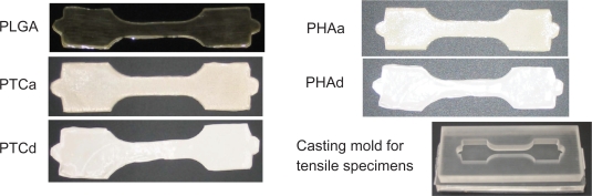
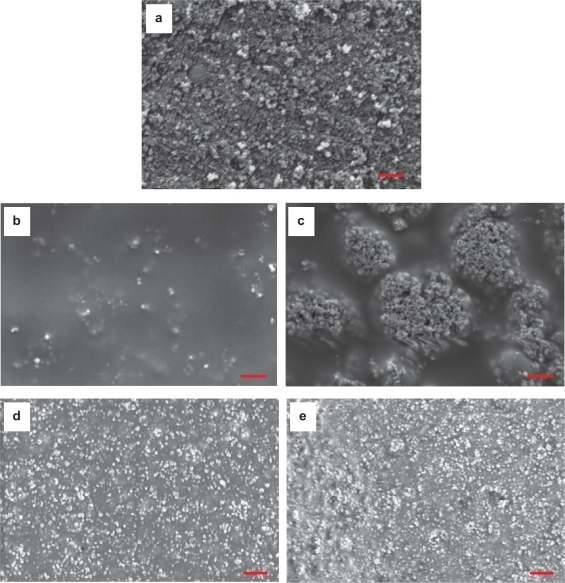
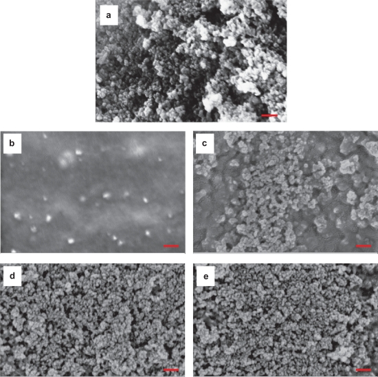
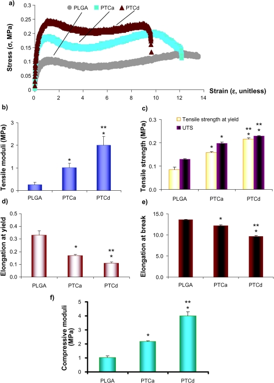
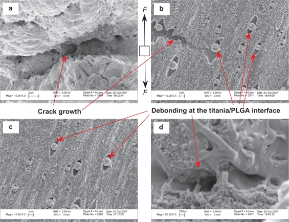
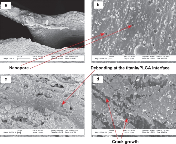
Ceramic/polymer composites have been considered as third-generation orthopedic biomaterials due to their ability to closely match properties (such as surface, chemistry, biological, and mechanical) of natural bone. It has already been shown that the addition of nanophase compared with conventional (or micron-scale) ceramics to polymers enhances bone cell functions. However, in order to fully take advantage of the promising nanometer size effects that nanoceramics can provide when added to polymers, it is critical to uniformly disperse them in a polymer matrix. This is critical since ceramic nanoparticles inherently have a strong tendency to form larger agglomerates in a polymer matrix which may compromise their properties. Therefore, in this study, model ceramic nanoparticles, specifically titania and hydroxyapatite (HA), were dispersed in a model polymer (PLGA, poly-lactic-co-glycolic acid) using high-power ultrasonic energy. The mechanical properties of the resulting PLGA composites with well-dispersed ceramic (either titania or HA) nanoparticles were investigated and compared with composites with agglomerated ceramic nanoparticles. Results demonstrated that well-dispersed ceramic nanoparticles (titania or HA) in PLGA improved mechanical properties compared with agglomerated ceramic nanoparticles even though the weight percentage of the ceramics was the same. Specifically, well-dispersed nanoceramics in PLGA enhanced the tensile modulus, tensile strength at yield, ultimate tensile strength, and compressive modulus compared with the more agglomerated nanoceramics in PLGA. In summary, supplemented by previous studies that demonstrated greater osteoblast (bone-forming cell) functions on well-dispersed nanophase ceramics in polymers, the present study demonstrated that the combination of PLGA with well-dispersed nanoceramics enhanced mechanical properties necessary for load-bearing orthopedic/dental applications.
Keywords: PLGA; agglomeration; biodegradable polymer; ceramic nanoparticles; dispersion; hydroxyapatite nanoparticles; mechanical properties; nanocomposites; orthopedic/dental applications; titania nanoparticles.
Figures
Similar articles
-
Altered responses of chondrocytes to nanophase PLGA/nanophase titania composites.Biomaterials. 2004 Mar-Apr;25(7-8):1205-13. doi: 10.1016/j.biomaterials.2003.08.012. Biomaterials. 2004. PMID: 14643594
-
Mechanisms of greater cardiomyocyte functions on conductive nanoengineered composites for cardiovascular application.Int J Nanomedicine. 2012;7:5653-69. doi: 10.2147/IJN.S34574. Epub 2012 Nov 13. Int J Nanomedicine. 2012. PMID: 23180962 Free PMC article.
-
Effect of ceramic filler content on the mechanical and thermal behaviour of poly-L-lactic acid and poly-L-lactic-co-glycolic acid composites for medical applications.J Mater Sci Mater Med. 2010 Sep;21(9):2523-31. doi: 10.1007/s10856-010-4110-9. Epub 2010 Jun 15. J Mater Sci Mater Med. 2010. PMID: 20552389
-
Polymeric nanocomposites: compounding and performance.J Nanosci Nanotechnol. 2008 Apr;8(4):1582-96. J Nanosci Nanotechnol. 2008. PMID: 18572559 Review.
-
Controlled arrangement of nanoparticle arrays in block-copolymer domains.Small. 2006 May;2(5):600-11. doi: 10.1002/smll.200500474. Small. 2006. PMID: 17193094 Review.
Cited by
-
In Vivo Degradation Studies of PGA-PLA Block Copolymer and Their Histochemical Analysis for Spinal-Fixing Application.Polymers (Basel). 2022 Aug 16;14(16):3322. doi: 10.3390/polym14163322. Polymers (Basel). 2022. PMID: 36015579 Free PMC article.
-
Biodegradation and cytotoxicity of ciprofloxacin-loaded hydroxyapatite-polycaprolactone nanocomposite film for sustainable bone implants.Int J Nanomedicine. 2015 Oct 1;10 Suppl 1(Suppl 1):119-27. doi: 10.2147/IJN.S79995. eCollection 2015. Int J Nanomedicine. 2015. PMID: 26491313 Free PMC article.
-
Nanophase hydroxyapatite and poly(lactide-co-glycolide) composites promote human mesenchymal stem cell adhesion and osteogenic differentiation in vitro.J Mater Sci Mater Med. 2012 Oct;23(10):2543-52. doi: 10.1007/s10856-012-4709-0. Epub 2012 Jul 7. J Mater Sci Mater Med. 2012. PMID: 22772475
-
Development of titanium dioxide nanowire incorporated poly(vinylidene fluoride-trifluoroethylene) scaffolds for bone tissue engineering applications.J Mater Sci Mater Med. 2019 Aug 14;30(8):96. doi: 10.1007/s10856-019-6300-4. J Mater Sci Mater Med. 2019. PMID: 31414231 Free PMC article.
-
Participation of Polymer Materials in the Structure of Piezoelectric Composites.Polymers (Basel). 2024 Dec 23;16(24):3603. doi: 10.3390/polym16243603. Polymers (Basel). 2024. PMID: 39771453 Free PMC article. Review.
References
-
- Traykova T, Aparicio C, Ginebra MP, Planell JA. Bioceramics as nanomaterials. Nanomedicine. 2006;1(1):91–106. - PubMed
-
- Boccaccini AR, Maquet V. Bioresorbable and bioactive polymer/bioglass® composites with tailored pore structure for tissue engineering applications. Composites Sci Technol. 2003;63(16):2417–2429.
-
- Hutmacher DW. Scaffolds in tissue engineering bone and cartilage. Biomaterials. 2000;21(24):2529–2543. - PubMed
-
- Thomson RC, Yaszemski MJ, Powers JM, Mikos AG. Hydroxyapatite fiber reinforced poly(α-hydroxy ester) foams for bone regeneration. Biomaterials. 1998;19(21):1935–1943. - PubMed
-
- Marra KG, Szem JW, Kumta PN, DiMilla PA, Weiss LE. In vitro analysis of biodegradable polymer blend/hydroxyapatite composites for bone tissue engineering. J Biomed Mater Res. 1999;47(3):324–335. - PubMed
Publication types
MeSH terms
Substances
LinkOut - more resources
Full Text Sources
Other Literature Sources
