Review
. 2010 Sep;120(3):403-17.
doi: 10.1007/s00401-010-0694-x. Epub 2010 May 13.

Progressive multifocal leukoencephalopathy and promyelocytic leukemia nuclear bodies: a review of clinical, neuropathological, and virological aspects of JC virus-induced demyelinating disease

Affiliations
Review

Progressive multifocal leukoencephalopathy and promyelocytic leukemia nuclear bodies: a review of clinical, neuropathological, and virological aspects of JC virus-induced demyelinating disease

Yukiko Shishido-Hara. Acta Neuropathol. 2010 Sep.

Abstract

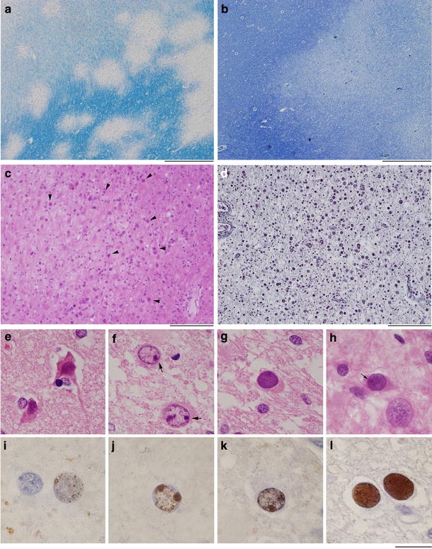
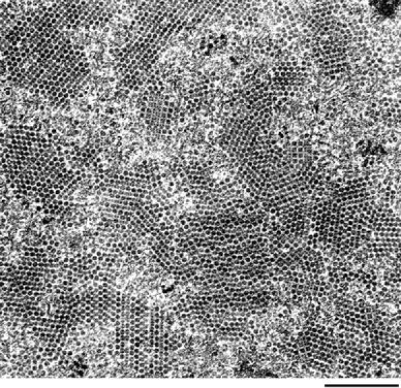
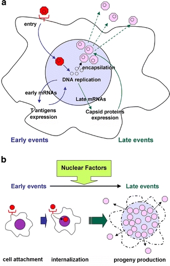
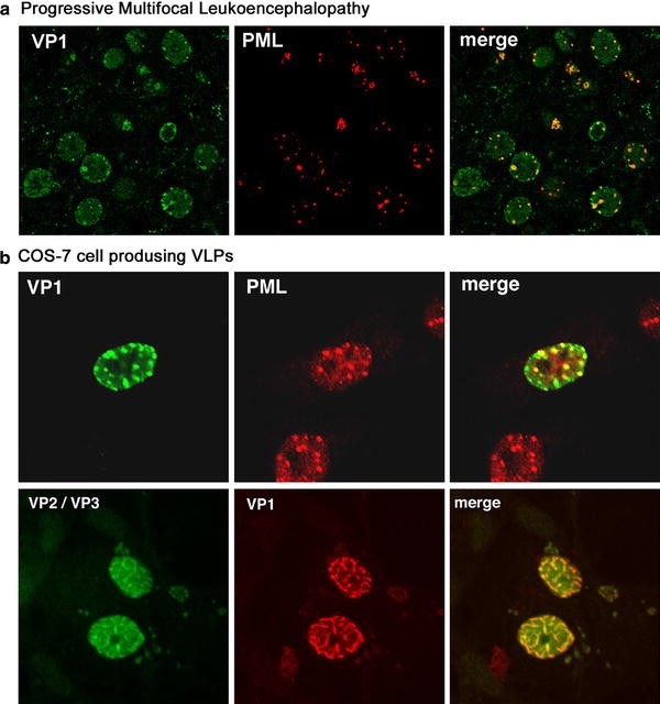
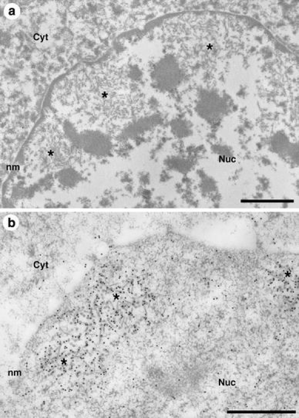
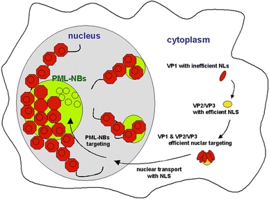
Progressive multifocal leukoencephalopathy is a fatal viral-induced demyelinating disease that was once rare but has become more prevalent today. Over the past decades, much has been learned about the disease from molecular study of the etiological agent of the disease, JC virus. Recently, promyelocytic leukemia nuclear bodies (PML-NBs), punctuate structures for important nuclear functions in eukaryotic cells, were identified as an intranuclear target of JC virus infection. Neuropathologically, JC virus-infected glial cells display diffuse amphophilic viral inclusions by hematoxylin-eosin staining (full inclusions), a diagnostic hallmark of this disease. Recent results using immunohistochemistry, however, revealed the presence of punctate viral inclusions preferentially located along the inner nuclear periphery (dot-shaped inclusions). Dot-shaped inclusions reflect the accumulation of viral progeny at PML-NBs, which may be disrupted after viral replication. Structural changes to PML-NBs have been reported for a variety of human diseases, including cancers and neurodegenerative disorders. Thus, PML-NBs may provide clues to the further pathogenesis of JC virus-induced demyelinating disease. Here, we review what we have learned since the disease entity establishment, including a look at recent progress in understanding the relationship between JC virus, etiology and PML-NBs.


Figures

Fig. 1
Fig. 1
Neuropathology of progressive multifocal leukoencephalopathy. a Characteristic demyelinating patterns, in which small well-demarcated lesions fuse to form larger lesions [Kluver-Barrera (KB) staining, bar 1.0 mm]. b Broad myelin pallor in the white matter (KB staining, bar 1.0 mm). c The same section shown in a. Glial cells harboring amphophilic inclusions in the enlarged nuclei (arrowheads) usually seen at the periphery of well-demarcated demyelinating lesions (HE staining, bar 100 μm). d The same section as in b. Glial cells with enlarged nuclei broadly scattered in the myelin pallor (Bodian staining, bar 100 μm). e Small and condensed nuclei of oligodendroglia surrounding cortex neurons. f Glial cells, potentially JC virus-infected oligodendroglias, display enlarged but clear nuclei without amphophilic inclusions. Dot-shaped structures (arrows) are detected and resemble dot-shaped JC viral inclusions shown in j and k. g, h Typical cells with intranuclear JC viral inclusions seen in the myelin pallor (g) and at the periphery of well-demarcated demyelination (h) (eh HE staining, bars 20 μm). i Immunohistochemistry (IHC) to detect the promyelocytic leukemia (PML) protein. Punctate PML distribution indicating the presence of PML-NBs. j, k Viral genomic DNA and capsid proteins VP2/VP3 are detected in punctate patterns (dot-shaped inclusions) by in situ hybridization (j) and IHC (k), respectively. l JC viral capsid proteins, by IHC, are detected throughout the nucleoplasm (full inclusions) (il bars 20 μm)
Fig. 2
Fig. 2
Electron micrograph of JC virions. JC virions, uniform in both size and shape display perfect alignment patterns called crystalloid arrays (Bar 1 μm). Courtesy of Drs. Shigeki Takeda and Hitoshi Takahashi
Fig. 3
Fig. 3
JC viral regulatory sequences. The viral regulatory sequences are divergent and divided into two groups, the “archetype” and “neurotropic type.” The archetype sequence consists of five sub-sequences, 25-, 23-, 55-, 66-, and 18-bp. The sequence of neurotropic types, such as Mad-1 and Tokyo-1, may be derived from the archetypal sequence via deletion and duplication. For details, refer to Yogo et al. [147]
Fig. 4
Fig. 4
The archetype hypothesis. a Archetypal viruses are frequently detected in the urine of healthy individuals, whereas neurotropic types are detected in the brains with progressive multifocal leukoencephalopathy. Thus, the archetypal viruses, which are ubiquitous in the human population, may occasionally transform into the neurotropic type, resulting in development of opportunistic demyelinating disease. b Detection of the archetype and neurotropic type in multiple tissues of carrier individuals has raised questions about the archetype hypothesis. See text for a detailed discussion
Fig. 5
Fig. 5
What determines JC virus neurotropism? a The viral life cycle begins with binding to cell surface receptors and is completed by the release of progeny from the host cell. Cellular susceptibility, for efficient production of viral progeny, can be regulated at any stage in the lytic cycle. b Although JC virus is now known to infect a variety of cells, the most efficient progeny production appears to occur in oligodendroglias, a phenomenon thought to be determined by multiple nuclear factors
Fig. 6
Fig. 6
Structures of JC virus late RNAs. Expression of major capsid protein VP1 and minor capsid proteins VP2/VP3 is kept roughly proportional by alternative splicing. VP1 is encoded on the M2 RNA, and VP2/VP3 on M1, downstream of the agnoprotein. Diagram based on Shishido-Hara et al. [116] (used with permission)
Fig. 7
Fig. 7
JC viral capsid proteins accumulate at PML-NBs in human brain tissue and COS-7 cells. JC virus capsid protein VP1 co-localizes in dots with the PML protein, a major component of PML-NBs. This was observed both in a human brain with progressive multifocal leukoencephalopathy (a) and in COS-7 cells producing VLPs (b top). Both VP1 and VP2/VP3 were distributed within the nucleus, associated with matrix-like structures (b bottom). a, b Top are from Shishido-Hara et al. [118] (used with permission)
Fig. 8
Fig. 8
Electron micrographs of JC virions in human brain tissues and of VLPs in COS-7 cells. a In human brain tissue with progressive multifocal leukoencephalopathy, electron microscopy reveals clustered native virions of both round and filamentous structures at the inner nuclear periphery, indicative of PML-NBs. b Consistently, in cultured cell lines producing VLPs, immunogold labeling VP1 epitopes is clustered at the nuclear periphery, where assembly of VLPs of both round and filamentous structures is detected (Bar 1 μm). Both panels are from Shishido-Hara et al. [117] (used with permission)
Fig. 9
Fig. 9
Schematic illustration of late events in the JC virus life cycle. VP1 may associate with VP2/VP3, and they are co-transported to the nucleus. Complexes of VP1 and VP2/VP3 are assembled into progeny virions at PML-NBs, and likely spread throughout the nucleoplasm, along with matrix-like structures
Fig. 10
Fig. 10
Does JC virus induce brain tumors in humans? a PML-NBs are the sites of multiple nuclear functions, and disruption or dysfunction of PML-NBs can cause a variety of human diseases, including leukemia and other cancers, as well as neurodegenerative disorders. Many viruses target PML-NBs. b JC virus-induced brain tumors are observed in experimental animals but whether or not this is clinically true in humans remains at issue

References

    1. Abdel-Aziz HO, Murai Y, Hong M, et al. Detection of the JC virus genome in lung cancers: possible role of the T-antigen in lung oncogenesis. Appl Immunohistochem Mol Morphol. 2007;15:394–400. doi: 10.1097/01.pai.0000213126.96590.64. - DOI - PubMed
    1. Angelini L, Pietrogrande MC, Delle Piane MR, et al. Progressive multifocal leukoencephalopathy in a child with hyperimmunoglobulin E recurrent infection syndrome and review of the literature. Neuropediatrics. 2001;32:250–255. doi: 10.1055/s-2001-19119. - DOI - PubMed
    1. Ariza A, Mate JL, Fernandez-Vasalo A, et al. p53 and proliferating cell nuclear antigen expression in JC virus-infected cells of progressive multifocal leukoencephalopathy. Hum Pathol. 1994;25:1341–1345. doi: 10.1016/0046-8177(94)90095-7. - DOI - PubMed
    1. Ariza A, Mate JL, Isamat M, Calatrava A, Fernandez-Vasalo A, Navas-Palacios JJ. Overexpression of Ki-67 and cyclins A and B1 in JC virus-infected cells of progressive multifocal leukoencephalopathy. J Neuropathol Exp Neurol. 1998;57:226–230. doi: 10.1097/00005072-199803000-00003. - DOI - PubMed
    1. Astrom KE, Mancall EL, Richardson EP., Jr Progressive multifocal leuko-encephalopathy; a hitherto unrecognized complication of chronic lymphatic leukaemia and Hodgkin’s disease. Brain. 1958;81:93–111. doi: 10.1093/brain/81.1.93. - DOI - PubMed

Publication types

MeSH terms