. 2010 Jul;5(4):617-31.
doi: 10.2217/rme.10.30.

Stem/progenitor cells from inflamed human dental pulp retain tissue regeneration potential

Affiliations

Stem/progenitor cells from inflamed human dental pulp retain tissue regeneration potential

Dominick J Alongi et al. Regen Med. 2010 Jul.

Abstract

Background: Potent stem/progenitor cells have been isolated from normal human dental pulps termed dental pulp stem cells (DPSCs). However, it is unknown whether these cells exist in inflamed pulps (IPs).

Aims: To determine whether DPSCs can be identified and isolated from IPs; and if they can be successfully cultured, whether they retain tissue regeneration potential in vivo.

Materials & methods: DPSCs from freshly collected normal pulps (NPs) and IPs were characterized in vitro and their tissue regeneration potential tested using an in vivo study model.

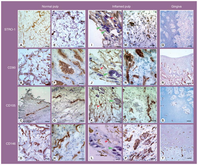
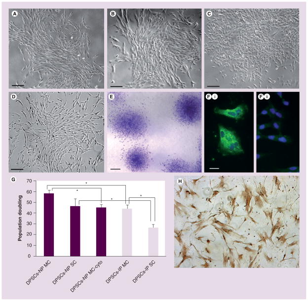
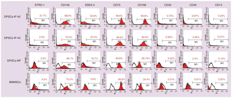
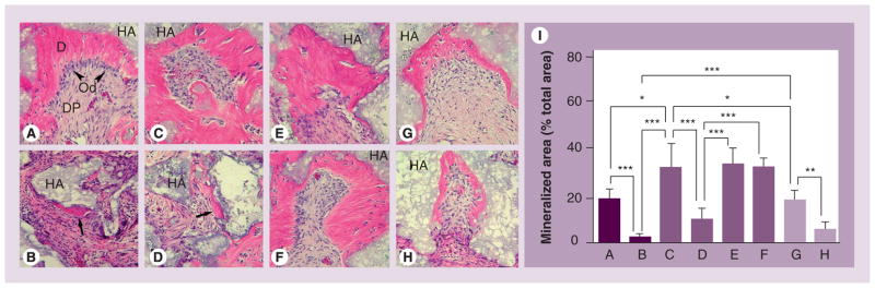
Results: The immunohistochemical analysis showed that IPs expressed higher levels of mesenchymal stem cell markers STRO-1, CD90, CD105 and CD146 compared with NPs (p < 0.05). Flow cytometry analysis showed that DPSCs from both NPs and IPs expressed moderate to high levels of CD146, stage-specific embryonic antigen-4, CD73 and CD166. Total population doubling of DPSCs-IPs (44.6 + or - 2.9) was lower than that of DPSCs-NPs (58.9 + or - 2.5) (p < 0.05), and DPSCs-IPs appeared to have a decreased osteo/dentinogenic potential compared with DPSCs-NPs based on the mineral deposition in cultures. Nonetheless, DPSCs-IPs formed pulp/dentin complexes similar to DPSCs-NPs when transplanted into immunocompromised mice.

Conclusion: DPSCs-IPs can be isolated and their mesenchymal stem cell marker profiles are similar to those from NPs. Although some stem cell properties of DPSCs-IPs were altered, cells from some samples remained potent in tissue regeneration in vivo.


Figures

Figure 1
Figure 1. Immunohistochemical analysis of mesenchymal stem cell markers STRO-1, CD90, CD105 and CD146 in normal pulps (A–H), inflamed pulps (I–P) and gingival tissues (Q–T)
Red arrowheads point at representative brown staining associated with vascular structures. Green arrowheads indicate the mineral particles accumulated in the lumen of blood vessels. In negative controls, no detectable stain was observed (not shown). Scale bars: (A–D, I–L & Q–T) 100 μm; (E–H & M–P) 30 μm.
Figure 2
Figure 2. Clonogenic dental pulp stem cells in cultures and population doubling studies
(A) DPSCs-NP #2 from a 9-year-old female, tooth #6 at passage 0. (B) DPSCs-IP #1 from a 17-year-old male, tooth #19 at passage 4. (C) DPSCs-IP #2 from a 19-year-old male, tooth #30 at passage 0. (D) DPSCs-IP #3 from a 15-year-old male, tooth #30 at passage 0. (E) DPSCs-IP #2 SC-5a from a 19-year-old male, tooth #30 at passage 2. Cell fixed and stained with toluidine blue. (Fi) STRO-1 fluorescence staining (green) and DAPI nuclear staining (blue) of seeded DPSCs-IP #3 MC from a 15-year-old male, tooth #30 at passage 2. (Fii) Nonimmuned isotype control of the same cells in (Fi). Scale bars: (A–D) 200 μm; (E) 1 mm; (F) 20 μm. (G) Relative population doublings between DPSCs-NPs, cytokine (TNF-α plus IL-1β) treated DPSCs-NPs and DPSC-IPs. *Significant difference (p ≤ 0.005); error bars: standard error of the mean. (H) β-galactosidase expression of senesced DPSCs at the end of population doubling studies (no β-galactosidase staining was observed at low passage of DPSCs, data not shown). DPSC: Dental pulp stem cell; IP: Inflamed pulp; MC: Multiple colony; NP: Normal pulp; SC: Single colony.
Figure 3
Figure 3. Immunophenotype analysis of human dental pulp stem cells and bone marrow-derived mesenchymal stem cells by flow cytometry
Multiple colony-derived cells (1 × 10⁵) at passage 3 were incubated with specific monoclonal antibodies against cell surface marker antigens STRO-1, CD73, CD146, CD166, SSEA-4, CD14, CD34 and CD45 followed by secondary antibodies (R-phycoerythrin). Positive signals are indicated by the red area. Subclass-matched control antibodies were used for the controls (white area). M1 represents fluorescent intensity exceeding 99% of the controls. BMMSCs were used for comparison. DPSCs-IPs sample #2: DPSCs-IP from a 19-year-old male, tooth #30; DPSCs-IPs sample #3: DPSCs-IP from a 15-year-old male, tooth #30. BMMSC: Bone marrow-derived mesenchymal stem cell; DPSC: Dental pulp stem cell; FI: Fluorescence intensity; IP: Inflamed pulp; NP: Normal pulp; SSEA: Stage-specific embryonic antigen.
Figure 4
Figure 4. Differentiation induction analysis
DPSC-NP or DPSC-IP cultures from different samples indicated by numbers stimulated with induction media with or without the presence of cytokines for 2 weeks. Cultures in the wells stained with Alizarin red (A) or cells harvested for real-time reverse transcription-PCR analysis (B). The bar charts represent the relative expression levels of each gene normalized against GAPDH. DPSCs-NPs (n = 3), DPSCs-IPs (n = 2); error bars: mean ± standard error of the mean. Images under the adipo real-time PCR bar chart panel are representative data of Oil Red O staining of a few DPSCs-NPs with minimal oil droplet accumulation intracellularly after 6 weeks of adipogenic stimulation. No Oil Red O stain was observed in stimulated DPSC-IP cultures. Scale bar: 25 μm. ALP: Alkaline phosphatase; BSP: Bone sialoprotein; CBFA1/RUNX2: Core-binding factor-α1; CNPase: 2′3′ cyclic nucleotide 3′ phosphodiesterase; Cyto: TNF-α plus IL-1β presence in cultures during the entire incubation periods; Diff: Differentiation induction; DPSC: Dental pulp stem cell; DSPP: Dentin sialophosphoprotein; GAPDH: Glyceraldehyde-3-phosphate dehydrogenase; IP: Inflamed pulp; LPL: Lipoprotein lipase; NFM: Neurofilament M; NP: Normal pulp; OCN: Osteocalcin; PPARγ2: Peroxisome proliferating activated receptor γ2.
Figure 5
Figure 5. In vivo formation of pulp/dentin complexes by transplanted dental pulp stem cells
Cells mixed with HA/tricalcium phosphate were subcutaneously transplanted into Begie XIDIII nu/nu mice. After 8 weeks, the resected tissues were processed for hematoxylin and eosin staining. (A) Dental pulp stem cells (DPSCs) from normal pulps as the control. (B) DPSCs from inflamed pulp (DPSCs-IP) multiple colony (MC), sample #1, 17-year-old male tooth #19. (C–F) DPSCs-IP, sample #2, 19-year-old male tooth #30 ((C) MC, (D) SC-4a, (E) SC-5a and (F) SC-6b). (G & H) DPSCs-IP sample #3, 15-year-old male tooth #30 ((G) MC and (H) SC-6a). (I) Analysis of the amount of dentin-like mineralized tissues formed by DPSCs between different samples. The labels of the x-axis (A–H) correspond to the (A–H) images. Arrows: mineralized tissue; arrowheads: odontoblast-like cells. *p < 0.05; **p < 0.01; ***p < 0.005; error bars, standard error of the mean; n = 7. D: Dentin-like; DP: Dental pulp-like; HA: Hydroxyapatite; Od: Odontoblast-like.

Similar articles

Cited by

References

    1. Reffelmann T, Könemann S, Kloner RA. Promise of blood- and bone marrow-derived stem cell transplantation for functional cardiac repair: putting it in perspective with existing therapy. J Am Coll Cardiol. 2009;53:305–308. - PubMed
    1. Meirelles Lda S, Nardi NB. Methodology, biology and clinical applications of mesenchymal stem cells. Front Biosci. 2009;14:4281–4298. - PubMed
    1. Mobasheri A, Csaki C, Clutterbuck AL, Rahmanzadeh M, Shakibaei M. Mesenchymal stem cells in connective tissue engineering and regenerative medicine: applications in cartilage repair and osteoarthritis therapy. Histol Histopathol. 2009;24:347–366. - PubMed
    1. Huang GTJ, Sonoyama W, Liu Y, Liu H, Wang S, Shi S. The hidden treasure in apical papilla: the potential role in pulp/dentin regeneration and bioroot engineering. J Endod. 2008;34:645–651. - PMC - PubMed
    1. García-Castro J, Trigueros C, Madrenas J, Pérez-Simón JA, Rodriguez R, Menendez P. Mesenchymal stem cells and their use as cell replacement therapy and disease modelling tool. J Cell Mol Med. 2008;12:2552–2565. - PMC - PubMed

Publication types

MeSH terms