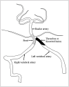
Lessons from the diagnosis and treatment of spontaneous vertebral arterial dissection. Case report
- PMID: 20465900
- PMCID: PMC3299023
- DOI: 10.1177/159101990901500211
Lessons from the diagnosis and treatment of spontaneous vertebral arterial dissection. Case report
Abstract
A 36-year-old man presented a sudden left occipital headache and right limb weakness after tooth-brushing. Conventional catheter digital subtraction angiography (DSA) showed a left VA occlusion at the crotch of the posterior inferior cerebellar artery. Four days later, the patient got worse. The angiogram showed the left vertebral artery had reopened and the basilar trunk occluded above the AICA. He died two days later and autopsy demonstrated a dissection of the basilar arteries. Based on the autopsy data from the patient in this study, we suggest that the BA dissection might be due to left VA dissection, and placing a stent on the juncture between the uninjured VA and the basilar trunk might be an effective method to prevent fatal BA occlusion.
Figures


Similar articles
-
Endovascular therapy for acute basilar artery occlusion caused by vertebral artery dissection: Case report.Medicine (Baltimore). 2021 Nov 24;100(47):e27995. doi: 10.1097/MD.0000000000027995. Medicine (Baltimore). 2021. PMID: 34964795 Free PMC article.
-
Endovascular treatment of huge dissecting aneurysms involving the basilar artery. Experience and lessons from two cases.Interv Neuroradiol. 2007 Dec;13(4):369-80. doi: 10.1177/159101990701300408. Epub 2008 Feb 1. Interv Neuroradiol. 2007. PMID: 20566106 Free PMC article.
-
Occipital Artery to Anterior Inferior Cerebellar Artery (AICA) Bypass for Treatment of a Ruptured Proximal AICA-Posterior Inferior Cerebellar Artery Mycotic Aneurysm.World Neurosurg. 2020 Oct;142:176-178. doi: 10.1016/j.wneu.2020.06.090. Epub 2020 Jun 23. World Neurosurg. 2020. PMID: 32585380
-
Childhood acute basilar artery thrombosis successfully treated with mechanical thrombectomy using stent retrievers: case report and review of the literature.Childs Nerv Syst. 2017 Feb;33(2):349-355. doi: 10.1007/s00381-016-3259-z. Epub 2016 Oct 4. Childs Nerv Syst. 2017. PMID: 27704247 Review.
-
[A Case of Fibromuscular Dysplasia in a Patient with Various Main Trunk Dissections in the Head and Neck over a Short Period].No Shinkei Geka. 2016 Jul;44(7):583-90. doi: 10.11477/mf.1436203334. No Shinkei Geka. 2016. PMID: 27384119 Review. Japanese.
Cited by
-
Three cases of Spontaneous Vertebral Artery Dissection (SVAD), resulting in two cases of Wallenberg syndrome and one case of Foville syndrome in young, healthy men.BMJ Case Rep. 2014 Apr 28;2014:bcr2014203945. doi: 10.1136/bcr-2014-203945. BMJ Case Rep. 2014. PMID: 24777086 Free PMC article.
-
Spontaneous Bilateral Vertebral Artery Dissection During a Basketball Game: A Case Report.Sports Health. 2016 Jan-Feb;8(1):53-6. doi: 10.1177/1941738114547347. Epub 2014 Aug 21. Sports Health. 2016. PMID: 26733592 Free PMC article.
References
-
- Caplan LR, Baquis GD, et al. Dissection of the intracranial vertebral artery. Neurology. 1998;38:868–877. - PubMed
-
- Hosoya T, Adachi M, et al. Clinical and neuroradiological features of intracranial vertebrobasilar artery dissection. Stroke; a journal of cerebral circulation. 1999;30:1083–1090. - PubMed
-
- Sasaki O, Ogawa H, et al. A clinicopathological study of dissecting aneurysms of the intracranial vertebral artery. Journal of Neurosurgery. 1991;75:874–882. - PubMed
-
- Schievink WI. Spontaneous dissection of the carotid and vertebral arteries. The New England Journal of Medicine. 2001;344:898–906. - PubMed
-
- Braverman IB, River Y, Rappaport JM, et al. Spontaneous vertebral artery dissection mimicking acute vertigo. Ann Otol Rhinol Laryngol. 1999;108:1170–1173. - PubMed
LinkOut - more resources
Full Text Sources

