. 2011 Apr;274(1-2):61-74.
doi: 10.1016/j.heares.2010.03.093. Epub 2010 May 11.

The medial geniculate, not the amygdala, as the root of auditory fear conditioning

Affiliations

The medial geniculate, not the amygdala, as the root of auditory fear conditioning

Norman M Weinberger. Hear Res. 2011 Apr.

Abstract

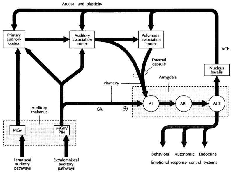
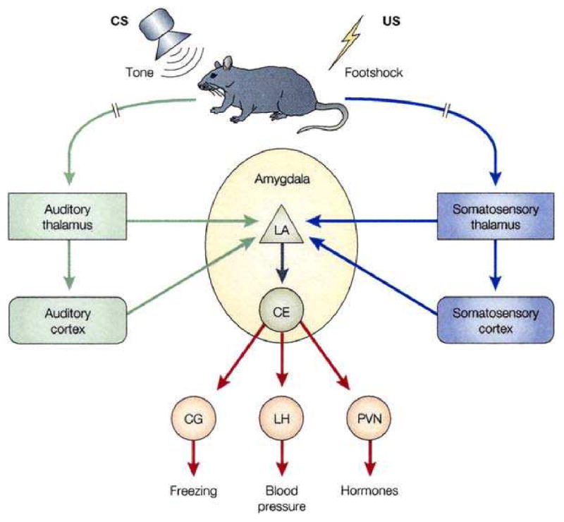
The neural basis of auditory fear conditioning (AFC) is almost universally believed to be the amygdala, where auditory fear memories are reputedly acquired and stored. This widely-accepted amygdala model holds that the auditory conditioned stimulus (CS) and the nociceptive unconditioned stimulus (US) first converge in the lateral nucleus of the amygdala (AL), and are projected independently to it from the medial division of the medial geniculate nucleus (MGm) and the adjacent posterior intralaminar nucleus (PIN), which serve merely as sensory relays. However, the four criteria that are used to support the AL model, (a) CS-US convergence, (b) associative plasticity, (c) LTP and (d) lesion-induced learning impairment, are also met by the MGm/PIN. Synaptic and molecular approaches supporting the AL also implicate the MGm/PIN. As both the AL and its preceding MGm/PIN are critically involved, we propose that the latter be considered the "root" of AFC.


Figures

Fig. 1
Fig. 1
A diagram illustrating some of the pathways underlying emotional information processing and response control by the amygdala. Pathways through which auditory inputs are transmitted to the amygdala are shown but similar circuits also exist for other sensory systems. Tonotopically organized auditory signals are transmitted to the auditory thalamus over lemniscal pathways, which synapse in the ventral division of the medial geniculate body (MGv). Extralemniscal pathways transmit to other parts of the auditory thalamus, including the medial division of the medial geniculate body (MGm) and the posterior intralaminar nucleus (PIN). While MGv only projects to primary auditory cortex, MGm/PIN projects to both primary and association areas of auditory cortex, as well as to the lateral nucleus of the amygdala (AL). The thalamo-amygdala projection forms asymmetric, excitatory (+) contacts with AL, contains glutamate (Glu), and may use this excitatory substance as a neurotransmitter. Thalamo-amygdala projections have been implicated in emotional learning, and high-frequency stimulation of these projections produces long-term potentiation (LTP) in AL. Auditory and polymodal association areas relay auditory signals to AL by way of the external capsule. These pathways are also involved in emotional learning and exhibit LTP. AL projects to the basolateral nucleus of the amygdala (ABL), which projects widely to cortical areas (not shown) and to the central nucleus of the amygdala (ACE). ACE has extensive connectivity with brainstem areas involved in the control of emotional responses. It also projects to the nucleus basalis, which projects widely to cortical areas. The pathway from the nucleus basalis to cortex uses acetylcholine (ACh) as a neurotransmitter. Cholinergic transmission to the cortex from the nucleus basalis has been implicated in cortical arousal and plasticity. (LeDoux, 1992)
Fig. 2
Fig. 2
Neural pathways underlying fear conditioning. Fear conditioning is a procedure in which an emotionally neutral conditioned stimulus (CS) is presented in association with an aversive unconditioned stimulus (US). In studies of rats, the CS has typically been an auditory tone and the US an electric footshock. The pathways mediating auditory fear conditioning in rats involve the convergence of the CS and US pathways onto single cells in the lateral nucleus of the amygdala (LA) from thalamic and cortical processing regions in the sensory systems that process the CS (auditory system) and US (somatosensory system). The LA then connects with the CE both directly and by way of other amygdala regions (not shown). Outputs of the CE then control the expression of fear responses, including freezing behavior and related autonomic nervous system (e.g., blood pressure and heart rate) and endocrine (pituitary–adrenal hormones) responses. Lesion and imaging studies, described in the text, have confirmed that the human amygdala is also involved in fear conditioning, but the involvement of subregions of the amygdala is still poorly understood in humans. CG, central gray; LH, lateral hypothalamus; PVN, paraventricular hypothalamus. From Medina et al. (2002). (Phelps and LeDoux, 2005)
Fig. 3
Fig. 3
Typical distribution of principal neuron types at the junction of the anterior and middle thirds of the medial geniculate body. Transverse section, Golgi–Cox. 15-day-old cat. (Morest, 1964)
Fig. 4
Fig. 4
Heterosynaptic long term potentiation (LTP) of auditory stimuli by the MGm. Stimulation of the MGm (200 Hz, 50 ms) paired with a preceding click produces LTP of click evoked local field potentials (LFPs) in the primary auditory cortex. Group mean (± SE) before and for 2 h after 30 click–stimulation pairings (n = 9). Probe clicks were given at 0.2 Hz before and for 60 min after pairing, and less often thereafter. Note the large magnitude of potentiation (ordinate Z-scores above pre-stimulation). Controls exhibited no facilitation. (Weinberger et al., 1995)
Fig. 5
Fig. 5
Stimulation of the posterior intralaminar nucleus (PIN), but not other medial geniculate nuclei, serves as an effective unconditioned stimulus (US) in auditory fear conditioning. (A) Behavioral conditioned bradycardia (“Condit”, n = 25) or lack thereof in a sensitization control group (“Sens.”, n = 9) to tone and shock. Guinea pigs received 10 trials of the CS and US unpaired followed by 30 trials of CS-US paired in the Conditioning group, while there was no change in protocol for the Sensitization control group. Note elicitation of novelty-induced bradycardia during the first five trials, which rapidly habituated by the end of the second block of unpaired presentation (block 2) for both groups. However, when pairing was initiated, the Conditioning group rapidly developed CS-elicited bradycardia during the first block of five trials, which continued to increase in magnitude during training (Edeline and Weinberger, 1992). (B) Substitution of stimulation of the PIN for a shock US produces conditioned bradycardia. However, stimulation of the ventral division (MGv), medial division (MGm) and dorsal/suprageniculate nuclei (MGd/SG, data combined) was ineffective and did not differ significantly from the Sensitization control group that received tone and shock unpaired (see [A] above). The ordinate is the magnitude of change in heart rate during the CS relative to pre-trial baseline. (Cruikshank et al., 1992)
Fig. 6
Fig. 6
Long term potentiation (LTP) of responses (local field potentials) of the MGm to brief high frequency (100 Hz, 285 ms) stimulation of its afferents from the brachium of the inferior colliculus (BIC). (A) The average amplitude of the MGm response, with respect to the baseline (n = 7). Note the growth in amplitude over the 1 h post-stimulation recording period. (B) The mean latency to peak of the local field potential. Note the decrease in latency over time. Vertical bars represent ± 1 standard error of the mean. (Gerren and Weinberger, 1983)
Fig. 7
Fig. 7
CS-specific receptive field plasticity in the medial division of the medial geniculate body in auditory fear conditioning. Guinea pigs received 30 trials of tone–shock pairing and developed behavior signs of AFC, i.e., conditioned bradycardia to the CS tone. (A) An example of frequency receptive fields before (“Pre”) and both immediately and 1 hour after (“Post”) the single conditioning session. (A1) Before conditioning, the best frequency was 2.5 kHz; the CS was selected to be 4.5 kHz. Immediately post-conditioning, there were increased responses at 4.0 and 4.5 kHz and some lower frequencies. (A2) RF difference function, i.e., the pre RF was subtracted from the post-training RF. Note that the maximum increase is at 4.5 kHz, the CS frequency. There was no change in response of the pre-training best frequency of 2.5 kHz. (A3) Comparable data obtained after a retention period of 1 h, i.e., with no additional training, shows continued development (“neural consolidation”). Compared to the pre-training RF, responses to some non-CS frequencies have declined, producing a shift in tuning so that the CS frequency became the new BF. (A4) The RF difference function shows that the largest increase in response is maintained at the CS frequency. In this case, the bandwidth (bw) of CS-related facilitation of response has been maintained as well. (B) MGm group data shown as normalized mean group RF difference functions for CS-specific plasticity. Graphs show percent change as a function of octave distance from the CS frequency. (B1) Immediately after conditioning (n = 16), the CS frequency showed a marked increase while frequencies as close as 0.1 octaves showed no change or decreased response. (B2) One hour later, the magnitude and specificity of CS-specific RF plasticity were maintained for cells still recorded (n = 10). CS-specific facilitation and CS-directed tuning shifts were never found in sensitization control subjects. (Edeline and Weinberger, 1992)

Similar articles

Cited by

References

    1. Aitkin LM. Medial geniculate body of the cat: Responses to tonal stimuli of neurons in medial division. J Neurophysiol. 1973;36(2):275–283. - PubMed
    1. Aitkin LM, Webster WR. Medial geniculate body of the cat: Organization and responses to tonal stimuli of neurons in ventral division. J Neurophysiol. 1972;35(3):365–380. - PubMed
    1. Amorapanth P, LeDoux JE, Nader K. Different lateral amygdala outputs mediate reactions and actions elicited by a fear-arousing stimulus. Nat Neurosci. 2000;3(1):74–79. - PubMed
    1. Andersen RA, Knight PL, Merzenich MM. The thalamocortical and corticothalamic connections of AI, AII, and the anterior auditory field (AAF) in the cat: Evidence for two largely segregated systems of connections. J Comp Neurol. 1980;194(3):663–701. - PubMed
    1. Apergis-Schoute AM, Debiec J, Doyère V, LeDoux JE, Schafe GE. Auditory fear conditioning and long-term potentiation in the lateral amygdala require ERK/MAP kinase signaling in the auditory thalamus: A role for presynaptic plasticity in the fear system. J Neurosci. 2005;25(24):5730–5739. - PMC - PubMed

Publication types