. 2010 Sep;12(10):716-26.
doi: 10.1016/j.micinf.2010.04.014. Epub 2010 May 11.

Protein kinase A catalytic subunit interacts and phosphorylates members of trans-sialidase super-family in Trypanosoma cruzi

Affiliations

Protein kinase A catalytic subunit interacts and phosphorylates members of trans-sialidase super-family in Trypanosoma cruzi

Yi Bao et al. Microbes Infect. 2010 Sep.

Abstract

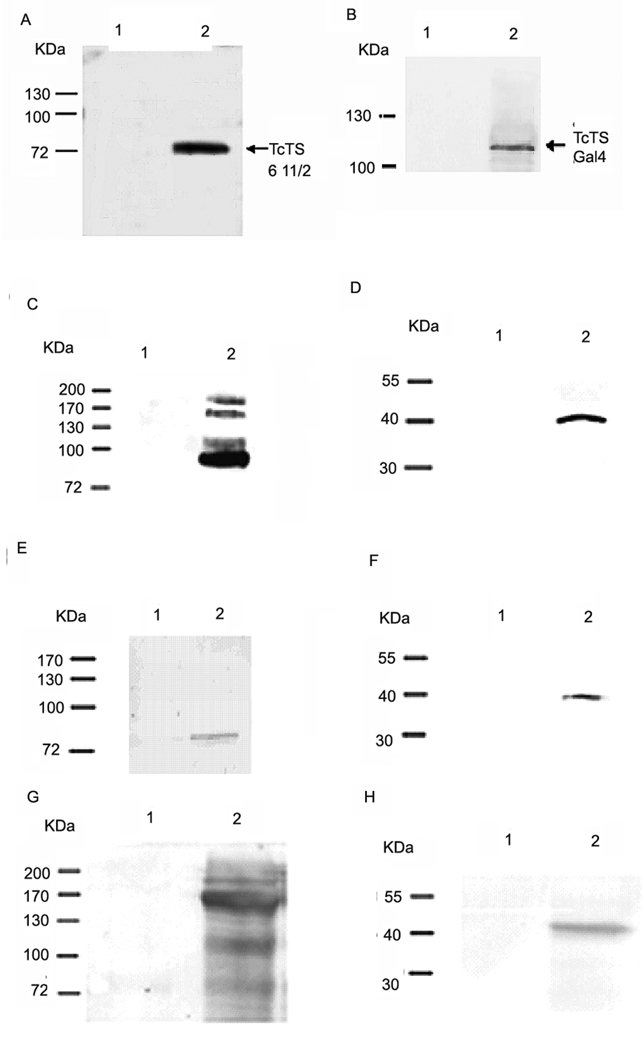
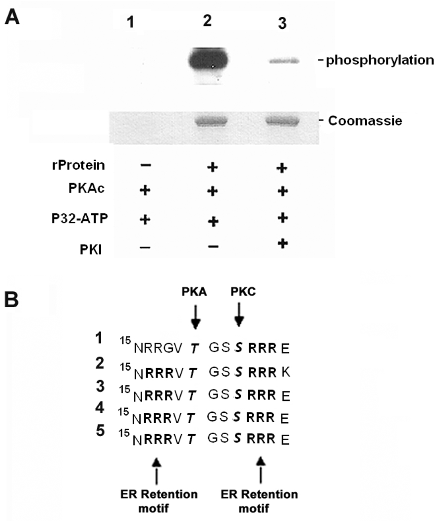
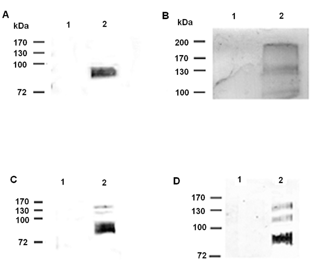
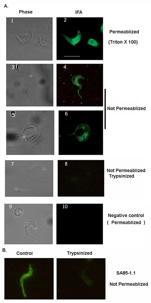
Protein kinase A (PKA) has been suggested as a regulator of stage differentiation in Trypanosoma cruzi. Using a yeast two-hybrid system we have begun to characterize the downstream substrates of T. cruzi PKA. We identified several members of the trans-sialidase super family by this approach. Immunoprecitation demonstrated that a TcPKAc monoclonal antibody was able to pull-down proteins recognized by trans-sialidase antibodies as well as a SA85-1.1 antibody and vice versa. An in vitro phosphorylation assay demonstrated that PKA phosphorylated the recombinant protein of an active trans-sialidase. In addition, a phospho-(Ser/Thr) PKA substrate antibody detected bands on immunoblot analysis of trans-sialidase antibody precipitated proteins from parasite lysate and the media of L(6)E(9) myoblasts infected with trypomastigotes as well as from a SA85-1.1 antibody precipitated proteins from parasite lysate. Immunofluorescence analysis suggested that some TcPKAc localizes to the plasma membrane surface of trypomastigotes. The identified trans-sialidases have PKA consensus phosphorylation sites located near the endoplasmic reticulum retention motif in the N-terminal. These data support that PKA phosphorylates trans-sialidase super family members in vivo.


Figures

Fig. 1
Fig. 1. T. cruzi trans-sialidase proteins interact with TcPKAc
Using a yeast two-hybrid system six members of the trans-sialidase super family target plasmids interacting with pBD- TcPKAc were obtained. Four ORFs of these genes and two partial genes were subcloned into a pAD-Gal4 vector. Each pAD-Gal4 construct was co-transformed into yeast with pBD-TcPKAc and cultured in high stringency condition (-Leu, -Trp, -His) (1) trans-sialidase Tc00. 1047053508563.20; (2) trans-sialidase Tc00.1047053509187.10; (3) trans-sialidase-like protein SA85-1.1 Genbank accession number X53545; (4) trans-sialidase Tc00.1047053507839.40; (5) trans-sialidase Tc00.104753510111.20; (6) trans-sialidase active clone lacking SAPA repeats TCTS 611/2. (7) Positive control (pADwt + pBDwt); (8) Negative control (pADwt + pLaminC).
Fig. 2
Fig. 2. Verification of a trans-sialidase antibody and Interactions of TcPKAc with the members of trans-sialidase super family
A commercial anti- sialidases antibody (rabbit) was first tested and verified for its ability to recognize T. cruzi trans-sialidases recombinant proteins, including a purified active trans-sialidase and trans-sialidase fusion proteins in crude yeast lysates (primary antibody dilution 1:1000; secondary antibody dilution 1: 5000) (Fig.2A–B). Co-immunoprecitation was performed using Triton X-100 protein extracts from trypomastigotes and a trans-sialidase polyclonal antibody (anti-TCTS 6 11/2, mouse), a commercial sialidase antibody, a T. cruzi SA85-1.1polyclonal antibody (rabbit) and a TcPKAc mAb(antibody dilutions:1;100 for immunoprecipitation, 1:1000 for immunoblot)(Fig.2C–H). A. The sialidase antibody binds to recombinant protein of TCTS 611/2. Lane1, 100ug of BSA as negative control; Lane 2, 100ng of recombinant protein of TCTS 611/2. B. The sialidase antibody binds to fusion protein of trans-sialidase (Tc00.1047053509187.10) in yeast lysate. Lane 1, 150 ug of wild type yeast lysate without transformation as negative control; Lane 2, 150ug of yeast lysate with transformation of both bait and pAD- trans-sialidase (Tc00.104053509187.10) constructs. Antibody recognized the pAD- trans-sialidase protein. C. Co-IP in Triton X −100 protein extracts from trypomastigotes with a TcPKAc mAb. Complex pulled down by a TcPKAc mAb contained proteins recognized by a commercial sialidase antibody on immunoblot. Lane 1, Negative control using an unrelated mAb (anti-bag5); Lane 2, Immunoprecipitation with TcPKAc mAb. There are four bands ranged from 85 kDa to 200 kDa, which reacted with this sialidase antibody. D. Co-IP in Triton X −100 protein extracts from trypomastigotes with a commercial sialidase antibody. The sialidase antibody was able to pull down a protein, which reacted with a TcPKAc mAb on immunoblot. Lane 1, negative control using pre-immune rabbit serum; Lane 2, Immunoprecipitation with Trans-sialidase antibody. There is a 40-kDa TcPKAc protein band. E. Co-IP in Triton X −100 protein extracts from trypomastigotes with a TcPKAc mAb. Complex pulled down by a TcPKAc mAb contained proteins recognized by a SA85-1.1 antibody on immunoblot. Lane 1, Negative control using an unrelated mAb (anti-bag5); Lane 2, Immunoprecipitation with TcPKAc mAb. There is an 85 kDa SA85-1.1 protein band. F. Co-IP in Triton X −100 protein extracts from trypomastigotes with a SA85-1.1 antibody. Precipitated complex by a SA85-1.1 antibody contained TcPKAc. Lane 1, Negative control using pre-immune rabbit serum; Lane 2, Immunoprecipitation with SA85-1.1 antibody. There is a 40-kDa TcPKAc protein band. G. Co-IP in Triton X −100 protein extracts from trypomastigotes with a TcPKAc mAb. Complex pulled down by a TcPKAc mAb contained proteins recognized by a trans-sialidase antibody (anti-TCTS 6 11/2) on immunoblot. Lane 1, Negative control using an unrelated mAb (anti-bag5); Lane 2, Immunoprecipitation with TcPKAc mAb. There are bands with molecular weights from 85–200 kDa. H. Co-IP in Triton X −100 protein extracts from trypomastigotes with a TCTS 6 11/2 antibody. Precipitated complex by a TCTS 6 11/2 antibody contained TcPKAc. Lane 1, Negative control using pre-immune mouse serum; Lane 2, Immunoprecipitation with Anti-TCTS 6 11/2;
Fig. 3
Fig. 3. PKA phosphorylates trans-sialidase recombinant protein in vitro PKAc phosphorylation sites were found to flank ER retention motifs in the members of trans-sialidase superfamily
A. In vitro phosphorylation of trans-sialidase recombinant protein by PKA is confirmed. Lane1, Absence of trans-sialidase recombinant protein as negative control; Lane 2, Trans-sialidase recombinant protein was phosphorylated in absence of PKI; Lane 3, PKI inhibited the phosphorylation; − absence of the reagent; + presence of the reagent (for details see MATERIALS AND METHODS). B. Five trans-sialidase protein sequences interacting with TcPKAc possess RXR (Arg-X-Arg) ER retention motifs in N-terminal. A PKAc phosphorylation site and a PKC phosphorylation site flank with these sites. It has been reported that phosphorylation by PKA and PKC in ER and /or Golgi at these sites suppresses ER retention and regulates surface delivery to membrane. The sequences were lined up at N-terminal position 15, RXR is a typical ER retention signal; RRXT or RXXT (position17–20) is a PKA phosphorylation site and SXR (position 23–25) is a PKC phosphorylation site. (1). Trans-sialidase Tc00.1047053508563.20; (2). Trans-sialidase Tc00.1047053509187.10; (3). Trans-sialidase Tc00.1047053507839.40; (4). Trans-sialidase Tc00.104753510111.20; (5). Trans-sialidase Tc00.1047053508285.60.
Fig. 4
Fig. 4. PKA phosphorylates the members of trans-sialidase super-family in vivo
Triton X −100 protein extracts from trypomastigotes and L6E9 myoblast culture media heavily infected with trypomastigotes were used for immunoprecipitation by antibodies to trans-sialidase family and then immunoblot by a phospho-(Ser/Thr) PKA substrate antibody (rabbit poly-clonal antibody) (dilutions: 1:100 for immunoprecipitation; 1:1000 for immunoblot). A. Co-IP in Triton X −100 protein extracts from trypomastigotes with a SA85-1.1 antibody. Precipitated complex by a SA85-1.1 antibody contained a broad band recognized by a phospho-(Ser/Thr) PKA substrate antibody. Lane 1, Negative control using pre-immune rabbit serum; Lane 2, Immunoprecipitation with SA85-1.1 antibody. B. Co-IP in Triton X −100 protein extracts from trypomastigotes with a TCTS 6 11/2 antibody. Precipitated complex by a TCTS 6 11/2 antibody contained three bands recognized by a phospho-(Ser/Thr) PKA substrate antibody Lane 1, Negative control using pre-immune mouse serum; Lane 2, Immunoprecipitation with TCTS 6 11/2 antibody C. Immunoprecipitation using L6E9 myoblast culture media heavily infected with trypomastigotes. A commercial sialidase antibody precipitated several proteins of trans-sialidases in the media with T. cruzi infected L6E9 myoblast. Lane 1, Uninfected L6E9 myoblast culture medium as negative control. Lane 2, Media from L6E9 myoblast heavily infected with trypomastigotes. Note that there were several bands precipitated by the sialidase antibody and these bands reacted with the same antibody on immunoblot, indicating that they were shed trans-sialidases. D. Co-IP using L6E9 myoblast culture media heavily infected with trypomastigotes with a commercial sialidase antibody followed by immunoblot with a phospho-(Ser/Thr) PKA substrate antibody. The shed trans-sialidases reacted with the phospho-(Ser/Thr) PKA substrate antibody, indicating that they were phosphorylated by TcPKAc before being shed into the media. Lane 1, Uninfected L6E9 myoblast culture medium as negative control. Lane 2, Medium from L6E9 myoblast heavily infected with trypomastigotes.
Fig. 5
Fig. 5. TcPKAc localizes both in cytoplasm and on membrane
A. IFA was performed in trypomastigotes. With treatment of Triton X-100 for permeabilization, TcPKAc staining is evident in the cytoplasm (1–2 panel). Without permeabilization (no Triton X-100), the antibody also recognized TcPKAc on the surface membrane and flagellum (3–6 panels). Trypsin treatment dramatically reduced surface staining with TcPKAc mAb (7–8 panels). Unrelated mAb (anti-bag5 of Toxoplasma gondii) was used as negative control (9–10 panels). Scale bars = 10 µm. B. SA85-1.1 rabbit polyclonal antibody was used to confirm the effect of trypsin treatment of trypomastigotes. Control IFA was performed on parasites without trypsin treatment. Trypsinized parasites were obtained by the process described in Material and Method. Note that Trypsin treatment dramatically reduced surface staining with the SA85-1.1 antibody.

Similar articles

Cited by

References

    1. Weiss LM, Wittner M, Coyle C, Shah S, Tanowitz HB. Parasitic infections of the nervous system and the eye in AIDS. Neurological Infections and Epidemiology. 1997;2:113–127.
    1. Ferreira MS. Infections by protozoa in immunocompromised hosts. Mem Inst Oswaldo Cruz. 2000;95 Suppl 1:159–162. - PubMed
    1. Lages-Silva E, Ramirez LE, Silva-Vergara M, Chiari E. Chagasic meningoencephalitis in a patient with acquired immunodeficiency syndrome, diagnosis, follow-up, and genetic characterization of Trypanosoma cruzi. Clin Inf Dis. 2002;34:118–123. - PubMed
    1. Sartori AM, Lopes MH, Benvenuti LA, Caramelli B, di Pietro A, Nunes EV, Ramirez LP, Shikanai-Yasuda MA. Reactivation of Chagas’ disease in a human immunodeficiency virus-infected patient leading to severe heart disease with a late positive direct microscopic examination of the blood. Am J Trop Med Hyg. 1998;59:784–786. - PubMed
    1. Sartori AM, Lopes MH, Caramelli B, Duarte MI, Pinto PL, Neto V, Amato Shikanai-Yasuda M. Simultaneous occurrence of acute myocarditis and reactivated Chagas’ disease in a patient with AIDS. Clin Inf Dis. 1995;21:1297–1299. - PubMed

Publication types

LinkOut - more resources