Review
. 2010:95:641-56.
doi: 10.1016/S0091-679X(10)95032-2.

Reconstitution and functional analysis of kinetochore subcomplexes

Affiliations
Review

Reconstitution and functional analysis of kinetochore subcomplexes

Daniel R Gestaut et al. Methods Cell Biol. 2010.

Abstract

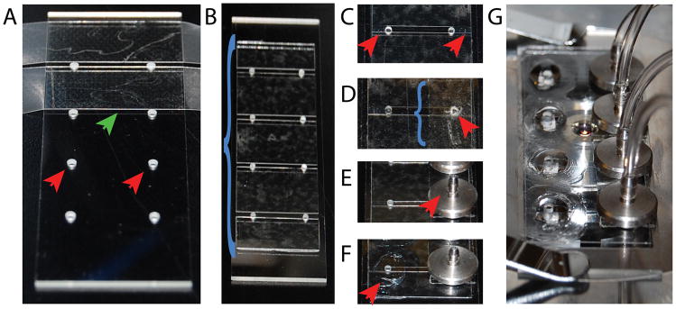
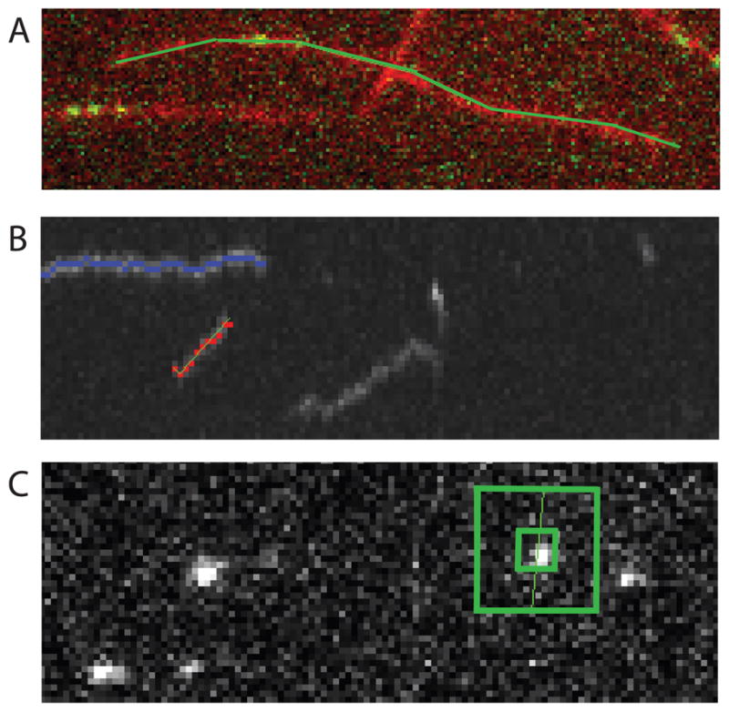
Kinetochores are multifunctional supercomplexes that link chromosomes to dynamic microtubule tips. Groups of proteins from the kinetochore are arranged into distinct subcomplexes that copurify under stringent conditions and cause similar phenotypes when mutated. By coexpressing all the components of a given subcomplex from a polycistronic plasmid in bacteria, many laboratories have had great success in purifying active subcomplexes. This has enabled the study of how the microtubule-binding subcomplexes of the kinetochore interact with both the microtubule lattice and dynamic microtubule tips. Here we outline methods for rapid cloning of polycistronic vectors for expression of kinetochore subcomplexes, their purification, and techniques for functional analysis using total internal reflection fluorescence microscopy (TIRFM).


Figures

Figure 1
Figure 1. Polycistronic cloning
(A) Schematic of the polycistronic vector developed by Tan et al. (B) Schematic of genes (multiple colors); 5′ primers containing Shine-Dalgarno sequence (green box), translational enhancer (blue box) and restriction enzyme recognition sequence; and 3′ primers with restriction enzyme recognition sequence for creating insertion products. (C) Schematic of final polycistronic vector.
Figure 2
Figure 2. TIRFM depth control
Schematic of the system that allows control of the depth of the TIRF field. In this system movement of the mirror will result in a displacement in the point of focus on the back focal plane. This movement ultimately results in a change in the depth of the TIRF field.
Figure 3
Figure 3. Simultaneous two-color imaging system
This system utilizes 2 dichroic mirrors to split and rejoin the two separately colored images side-by-side onto the same camera CCD. The images are refocused using separate lenses within each path.
Figure 4
Figure 4. Four Perfusion Chamber slide
(A) Glass slide with four holes (red arrowheads) drilled along each long axis, holes are spaced to allow double sided sticky tape (green arrowhead) to be placed between them. (B)One silanized coverslip (blue bracket) is placed onto the double sided sticky tape, and excess tape is removed. (C) Grease is used to seal the edges of the perfusion chambers (red arrowheads) up to the holes in the slide. (D) The slide is flipped over and adhesive transfertape (blue bracket) is placed above the holes along one long axis of the slide. A hole (red arrowhead) is created in the adhesive transfertape above the hole in the slide using forceps. (E) Custom flow adapters (red arrowhead) are attached to the slide via the adhesive transfertape. (F) Rings of grease (red arrowhead) are made around the holes opposite the flow adapters to create a pool for buffers. (G) Image of the final slide on the microscope with outlet tubing attached to the flow adapters.
Figure 5
Figure 5. TIRF data analysis
(A) Images from the red and green channels are overlapped and a multi-segmented line (green) is drawn along one of the microtubules. (B) Kymograph generated from step one, showing a preselected event (blue), currently selected event (green line, and red spots) and unselected events (grayscale). (C) Single image from the original tiff stack of the green channel (grayscale) in which a small box (green) is used to generate a brightness measurement, and a larger box (green) is used to generate a background measurement.

References

    1. Asbury CL, Gestaut DR, Powers AF, Franck AD, Davis TN. The Dam1 kinetochore complex harnesses microtubule dynamics to produce force and movement. Proc Natl Acad Sci U S A. 2006;103:9873–9878. - PMC - PubMed
    1. Berg HC. Random Walks in Biology. Princeton University Press; 1993. Revised edn.
    1. Cheeseman IM, Chappie JS, Wilson-Kubalek EM, Desai A. The conserved KMN network constitutes the core microtubule-binding site of the kinetochore. Cell. 2006;127:983–997. - PubMed
    1. Cheeseman IM, Desai A. Molecular architecture of the kinetochore-microtubule interface. Nat Rev Mol Cell Biol. 2008;9:33–46. - PubMed
    1. Cras JJ, Rowe-Taitt CA, Nivens D, Ligler FS. Comparison of chemical cleaning methods of glass in preparation for silanization. Biosensors and Bioelectronics. 1999;14:683–688.

MeSH terms

Substances