. 2010 Jul 1;9(7):785-95.
doi: 10.1016/j.dnarep.2010.04.002. Epub 2010 May 13.

Direct interaction between XRCC1 and UNG2 facilitates rapid repair of uracil in DNA by XRCC1 complexes

Affiliations

Direct interaction between XRCC1 and UNG2 facilitates rapid repair of uracil in DNA by XRCC1 complexes

Mansour Akbari et al. DNA Repair (Amst). .

Abstract

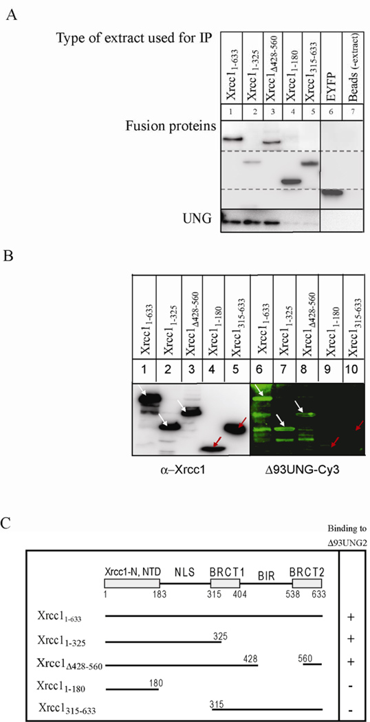
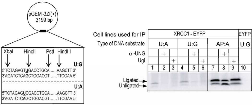
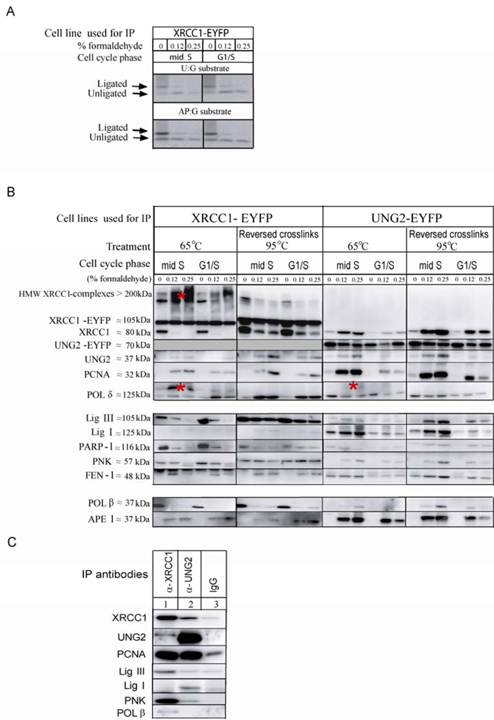
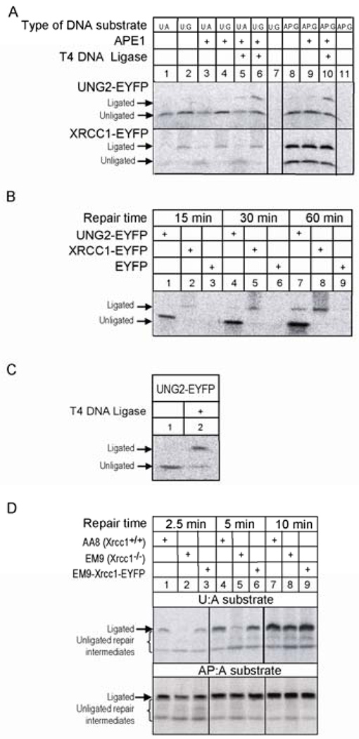
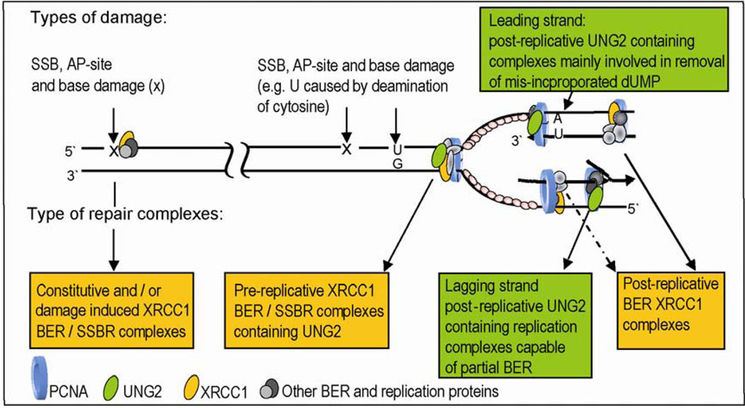
Uracil-DNA glycosylase, UNG2, interacts with PCNA and initiates post-replicative base excision repair (BER) of uracil in DNA. The DNA repair protein XRCC1 also co-localizes and physically interacts with PCNA. However, little is known about whether UNG2 and XRCC1 directly interact and participate in a same complex for repair of uracil in replication foci. Here, we examine localization pattern of these proteins in live and fixed cells and show that UNG2 and XRCC1 are likely in a common complex in replication foci. Using pull-down experiments we demonstrate that UNG2 directly interacts with the nuclear localization signal-region (NLS) of XRCC1. Western blot and functional analysis of immunoprecipitates from whole cell extracts prepared from S-phase enriched cells demonstrate the presence of XRCC1 complexes that contain UNG2 in addition to separate XRCC1 and UNG2 associated complexes with distinct repair features. XRCC1 complexes performed complete repair of uracil with higher efficacy than UNG2 complexes. Based on these results, we propose a model for a functional role of XRCC1 in replication associated BER of uracil.


Conflict of interest statement

Conflict of interest

The authors declare that there are no conflicts of interest.

Figures

Fig. 1
Fig. 1
Intracellular localisation of XRCC1 and UNG2. (A) Confocal microscopy analysis of co-localisation of XRCC1-ECFP, UNG2-EYFP and Hc-Red-PCNA in S-phase (upper row) and non-S-phase cells (lower row). (B) Immunohistochemistry. Upper row: HeLa cells expressing 3 × FLAG-UNG2-EYFP were fixed and incubated with a monoclonal α-FLAG antibody. Mid row: CHO EM9 cells were transiently tranfected with XRCC1-EYFP, fixed and stained with a polyclonal α-XRCC1 antibody. Lower row: HeLa cells expressing 3 × FLAG-UNG2-EYFP were fixed and incubated with a monoclonal α-FLAG antibody and a polyclonal α-XRCC1 antibody. Detection was perfomed using Alexa Flour (532 and 647) labelled secondary antibodies. Arrows show non-tranfected cells that did not stain with the antibodies. (C) In situ Proximity Ligation Assay (PLA). Upper row: HeLa cells expressing 3 × FLAG-UNG2-EYFP (UNG2-EYFP) were fixed and incubated with a monoclonal α-FLAG antibody and a polyclonal α-XRCC1 antibody followed by detection of PLA-foci using oligonucleotide conjugated probe antibodies directed against the primary antibodies. Annealing of the probes occurs when the target proteins are in close proximity, which initiates the amplification of a reporter signal and appears as foci (PLA, α-Flag and α-XRCC1). An overview of several cells after PLA (Overview, PLA)(far left, the nuclei are enclosed) and a merged image of PLA and UNG2-EYFP are shown (Merged)(far right). Lower row: same as upper row but the cells were not stained with the α-Flag antibody (PLA ctr). Bar, 5 µm.
Fig. 2
Fig. 2
Pull-down and Far Western analysis of Xrcc1 and UNG2 interaction. The indicated constructs of Cricetulus griseus Xrcc1 fused with ECFP were transiently expressed in Xrcc1 deficient Chinese hamster ovary cell line EM9. Cells expressing only EYFP were used as control. The fusion proteins were immunoprecipitated with paramagnetic beads covalently coupled with α-GFP. (A) The immunoprecipitates were incubated with the catalytic domain of UNG2 (Δ93UNG2). The beads were suspended in loading buffer and subjected to Western analysis. Lane 7, the beads were only incubated with recombinant UNG. The membrane was incubated with α-GFP antibody (upper panel) and α-UNG antibody (lower panel). (B) Far Western analysis. The immunoprecipitates were separated on gel and transferred to a PDF membrane and refolded. The membrane was first incubated with Cy-3 labelled Δ93UNG2 then stripped and probed with α-XRCC1. White arrows shows binding of Δ93UNG2 to XRCC1-fusion construct, while red arrows indicate no binding. (C) Schematic overview of the XRCC1 deletion constructs applied and their ability to bind to recombinant Δ93UNG2.
Fig. 3
Fig. 3
BER analysis of XRCC1-EYFP immunoprecipitates from whole cell extract. Left panel: schematic illustration of U:G and U:A DNA substrate for BER analysis. AP site DNA was prepared by incubation of uracil-containing DNA with recombinant UNG. Right panel: lanes 1 to 6, uracil-BER analysis of XRCC1-EYFP complexes in the absence or presence of UNG2 neutralising antibodies (α-UNG2) or Ugi. Lanes 7 to 9, analysis of AP site BER. Lane 10 shows BER analysis of the control immunoprecipitates from EYFP-expressing cells. The repair reaction was carried out at 32 °C for 15 min. The repaired DNA was digested with XbaI / HindIII to get information about the ligated and unligated repair products as shown.
Fig. 4
Fig. 4
Comparative Western blot analysis of XRCC1-EYFP and UNG2-EYFP immunoprecipitates from extracts of mid S- and G1/S-phase enriched cells. (A) BER activity of immunoprecipitated XRCC1-EYFP from crosslinked and non-crosslinked cell extracts. BER was carried out at 32 °C for 15 min. The repaired DNA was digested with XbaI and HindIII to get information about the ligated and unligated repair products. (B) Western analysis of immunoprecipitates from formaldehyde crosslinked and non-crosslinked XRCC1-EYFP and UNG2-EYFP expressing HeLa cell extracts. Cells were treated with formaldehyde (0, 0.125 or 0.25%) prior to whole cell extract preparation. Western blot analysis of the immunoprecipitates before (65 °C) and after (95 °C) reversal of the crosslinks is shown. (C) Western analysis of immunoprecipitates from insoluble chromatin-bound fraction of freely cycling HeLa cells using anti-XRCC1 (lane 1) or anti-UNG (lane 2) antibodies. One representative gel from three experiments is shown.
Fig. 5
Fig. 5
BER analysis of UNG2-EYFP and XRCC1-EYFP immunoprecipitates from mid S-phase enriched cells and BER analysis of cell extracts in the absence or presence of Xrcc1. (A) BER activity of the immunoprecipitated UNG2-EYFP and XRCC1-EYFP complexes was analyzed using U:A, U:G and AP:G DNA substrates with or without additional recombinant APE1 and T4 DNA ligase at 32 °C for 15 min. Lanes 7 and 11 show repair activity of the immunoprecipitates from EYFP-expressing cell extracts as control. (B) Time-course BER analysis of the immunoprecipitated UNG2-EYFP and XRCC1-EYFP complexes using U:G substrate. (C) End-trimming and DNA ligase activity of UNG2-EYFP immunoprecipitates. After 1 h incubation, half of the repaired DNA was further incubated with T4 DNA ligase (lane 2). (D) Time-course BER analysis of extracts from AA8 (Xrcc1+/+), EM9 (Xrcc1−/−) and EM9 cells expressing Xrcc1-EYFP (EM9-Xrcc1-EYFP) on a U:A and AP:A substrate. All DNA samples were digested with XbaI and HindIII to get information about DNA ligase and end-trimming activity of the immunoprecipitates.
Fig. 6
Fig. 6
A model for repair of uracil in DNA by XRCC1 and UNG2 complexes. UNG2 is linked to replication through its interaction with XRCC1 and PCNA, both of which interact with a number of proteins involved in DNA replication and repair. We suggest a model for how these interactions organize efficient pre-replicative repair of U:G mismatch and post-replicative repair of mis-incorporated uracil (U:A) in DNA, during replication.

References

    1. Lindahl T. Instability and decay of the primary structure of DNA. Nature. 1993;362:709–715. - PubMed
    1. Verri A, Mazzarello P, Biamonti G, Spadari S, Focher F. The specific binding of nuclear protein(s) to the cAMP responsive element (CRE) sequence (TGACGTCA) is reduced by the misincorporation of U and increased by the deamination of C. Nucleic Acids Res. 1990;18:5775–5780. - PMC - PubMed
    1. Viswanathan A, You HJ, Doetsch PW. Phenotypic change caused by transcriptional bypass of uracil in nondividing cells. Science. 1999;284:159–162. - PubMed
    1. Akbari M, Pena-Diaz J, Andersen S, Liabakk NB, Otterlei M, Krokan HE. Extracts of proliferating and non-proliferating human cells display different base excision pathways and repair fidelity. DNA Repair (Amst.) 2009;8:834–843. - PubMed
    1. Sanderson RJ, Mosbaugh DW. Fidelity and mutational specificity of uracil-initiated base excision DNA repair synthesis in human glioblastoma cell extracts. J. Biol. Chem. 1998;273:24822–24831. - PubMed

Publication types

LinkOut - more resources