. 2011 Jan;21(1):222-32.
doi: 10.1093/cercor/bhq083. Epub 2010 May 13.

Gonadectomy and hormone replacement affects in vivo basal extracellular dopamine levels in the prefrontal cortex but not motor cortex of adult male rats

Affiliations

Gonadectomy and hormone replacement affects in vivo basal extracellular dopamine levels in the prefrontal cortex but not motor cortex of adult male rats

T Aubele et al. Cereb Cortex. 2011 Jan.

Abstract

Gonadectomy in adult male rats is known to impair performance on dopamine (DA)-dependent prefrontal cortical tasks and selectively dysregulate end points in the mesoprefrontal DA system including axon density. In this study, in vivo microdialysis and high-pressure liquid chromatography were used to determine whether short (4 day)- and/or long-term (28 day) gonadectomy and hormone replacement might also influence the more functionally relevant metric of basal extracellular DA level/tone. Assessments in medial prefrontal cortex revealed that DA levels were significantly lower than control in 4-day gonadectomized rats and similar to control in 4-day gonadectomized animals supplemented with both testosterone and estradiol. Among the long-term treatment groups, DA levels were significantly higher than control in gonadectomized rats and gonadectomized rats given estradiol but were similar to control in rats given testosterone. In contrast, extracellular DA levels measured in motor cortex were unaffected by long- or short-term gonadectomy. The effects of gonadectomy and hormone replacement on prefrontal cortical DA levels observed here parallel previously identified effects on prefrontal DA axon density and could represent hormone actions relevant to the modulation of DA-dependent prefrontal cortical function and perhaps its dysfunction in disorders such as schizophrenia, attention deficit hyperactivity disorder, and autism where males are disproportionately affected relative to females.


Figures
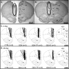
Figure 1.
Figure 1.
Representative serial photomicrographs (A,B) showing the placement of a microdialysis probe in the mPFC of a control animal. Boundaries between cytoarchitectonic fields and between cortex and white matter are marked with dashed lines in the unimplanted hemisphere and damage caused by the probe itself is circled in the other hemisphere. Line drawings in panels (C) and (D) are modified from Paxinos and Watson (1998) and illustrate the locations of microdialysis probe tracks (thick black lines) for each animal included in the 4-day (panel C) and 28-day (panel D) animal groups. For both the 4-day (C) and 28-day (D) cohorts, probes were comparably placed with respect to cortical cytoarchitecture (dashed lines) in rats that were sham operated (CTRL), gonadectomized (GDX), gonadectomized and supplemented with TP (GDX-TP), and gonadectomized and supplemented with E (GDX-E). The number that appears in parentheses below the drawings identifies the number of animal subjects in that group. Cg1, anterior cingulated cortex; Prl, Prelimbic cortex; IL, infralimbic cortex; DP, dorsopeduncular cortex; olf, olfactory bulb; wm, white matter.
Figure 2.
Figure 2.
Representative serial micrographs (A,B) showing the placement of a microdialysis probe in the motor cortex of a gonadectomized animal. Cytoarchitectonic fields and boundaries between cortex and white matter are marked with dashed lines in the unimplanted hemisphere, damage caused by the probe itself is circled in the other hemisphere, and the modest damage in the contralateral hemisphere caused by an anchoring screw is marked with an asterisk. Line drawings in panels (C) and (D) (modified from Paxinos and Watson [1998]) mark the locations of microdialysis probe tracks (thick black lines) for each animal in the 4-day (panel C) and 28-day (panel D) groups. For both the 4-day and 28-day cohorts, probes were comparably placed with respect to cortical cytoarchitecture (dashed lines) in rats that were sham operated (CTRL) and gonadectomized (GDX). The number that appears in parentheses below the drawings identifies the number of animal subjects in the group. Cg1, anterior cingulated cortex; Prl, prelimbic cortex; IL, infralimbic cortex; DP, dorsopeduncular cortex; olf, olfactory bulb; wm, white matter.
Figure 3.
Figure 3.
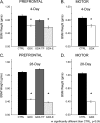
Bar graphs showing mean weights of the BSMs in grams (±standard error of the mean) for animals used in microdialysis assessments of mPFC (A,C) and motor cortex (B,D). For the PFC studies, data from animals that were sham operated (CTRL, black bars), gonadectomized (GDX, white bars), gonadectomized and supplemented with TP (GDX-TP, dark gray bars), or gonadectomized and supplemented with E (GDX-E, light gray bars) for 4 days (A) or for 28 days (C) are shown separately. For the motor cortex studies, data from animals that were sham operated (CTRL, black bars) or gonadectomized (GDX, white bars) for 4 days (B) or for 28 days (D) are also shown separately. Asterisks show that the mean weights of these androgen-sensitive muscles were significantly lower in all the 4- and 28-day GDX and GDX-E rats compared with the corresponding CTRL and GDX-TP groups.
Figure 4.
Figure 4.
Bar graphs showing mean extracellular DA level (fmol/μL) ± standard error of the mean in 4-day (A) and 28-day PFC (B) measured in animals that were sham operated (CTRL, black bars), gonadectomized (GDX, white bars), gonadectomized and supplemented with TP (GDX-TP, dark gray bars), or gonadectomized and supplemented with E (GDX-E, light gray bars). Mean DA level in the PFC was significantly lower in the GDX compared with CTRL rats and was similar to control in the GDX-TP and GDX-E groups in the 4-day animals. However, in the 28-day cohort, mean DA level in the PFC was significantly higher in the GDX and GDX-E compared with CTRL rats and was similar to CTRL in the GDX-TP group only. Regression plots that relate 20-min bin measurements of individual animals’ level of extracellular DA to the weights of their androgen-sensitive BSMs in grams (g) reveal no significant correlation between these 2 measures in 4-day rats (C) but that DA level and BSM weights are significantly correlated in 28-day animals (D). Open circles in the regression plots identify data points from GDX animals, and R2 values from the regression analyses appear in the upper left.
Figure 5.
Figure 5.
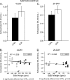
Bar graphs showing mean extracellular DA level (fmol/μL) ± standard error of the mean in 4-day (A) and 28-day motor cortex (B) measured in animals that were sham operated (CTRL, black bars), gonadectomized (GDX, white bars), for 28 days. There were no significant differences in extracellular DA level in motor cortex between the CTRL and GDX groups at either of these time points. Further, regression plots that relate 20-min bin sample measurements of individual animals’ level of extracellular DA to the weights of their androgen-sensitive BSMs in grams (g) reveal no significant correlations between these 2 variables for either the 4-day (C) or 28-day time point (D). Open circles in regression plots identify data points from GDX animals, and R2 values from the regression analyses appear in the upper left.

References

    1. Adler A, Vescovo P, Robinson JK, Kritzer MF. Gonadectomy in adult life increases tyrosine hydroxylase immunoreactivity in the prefrontal cortex and decreases open field activity in male rats. Neuroscience. 1999;89:939–954. - PubMed
    1. Akhondzadeh S, Rezaei F, Larijani B, Nejatisafa AA, Kashani L, Abbasi SH. Correlation between testosterone, gonadotropins and prolactin and severity of negative symptoms in male patients with chronic schizophrenia. Schizophr Res. 2006;84:405–410. - PubMed
    1. Aubele T, Kaufman R, Montalmant F, Kritzer MF. Effects of gonadectomy and hormone replacement on a spontaneous novel object recognition task in adult male rats. Horm Behav. 2008;54:244–252. - PMC - PubMed
    1. Balla A, Nattini ME, Sershen H, Lajtha A, Dunlop DS, Javitt DC. GABAB/NMDA receptor interaction in the regulation of extracellular dopamine levels in rodent prefrontal cortex and striatum. Neuropharmacology. 2009;56:915–921. - PMC - PubMed
    1. Barker GR, Bird F, Alexander V, Warburton EC. Recognition memory for objects, place, and temporal order: a disconnection analysis of the role of the medial prefrontal cortex and perirhinal cortex. J Neurosci. 2007;27:2948–2957. - PMC - PubMed

Publication types