. 2010 Aug 12;116(6):1002-10.
doi: 10.1182/blood-2010-02-269746. Epub 2010 May 13.

Complement inhibition decreases the procoagulant response and confers organ protection in a baboon model of Escherichia coli sepsis

Affiliations

Complement inhibition decreases the procoagulant response and confers organ protection in a baboon model of Escherichia coli sepsis

Robert Silasi-Mansat et al. Blood. .

Abstract

Severe sepsis leads to massive activation of coagulation and complement cascades that could contribute to multiple organ failure and death. To investigate the role of the complement and its crosstalk with the hemostatic system in the pathophysiology and therapeutics of sepsis, we have used a potent inhibitor (compstatin) administered early or late after Escherichia coli challenge in a baboon model of sepsis-induced multiple organ failure. Compstatin infusion inhibited sepsis-induced blood and tissue biomarkers of complement activation, reduced leucopenia and thrombocytopenia, and lowered the accumulation of macrophages and platelets in organs. Compstatin decreased the coagulopathic response by down-regulating tissue factor and PAI-1, diminished global blood coagulation markers (fibrinogen, fibrin-degradation products, APTT), and preserved the endothelial anticoagulant properties. Compstatin treatment also improved cardiac function and the biochemical markers of kidney and liver damage. Histologic analysis of vital organs collected from animals euthanized after 24 hours showed decreased microvascular thrombosis, improved vascular barrier function, and less leukocyte infiltration and cell death, all consistent with attenuated organ injury. We conclude that complement-coagulation interplay contributes to the progression of severe sepsis and blocking the harmful effects of complement activation products, especially during the organ failure stage of severe sepsis is a potentially important therapeutic strategy.


Figures

Figure 1
Figure 1
Complement activity and TCC antigen levels in plasma of baboons treated with compstatin during the first (T0) and second (T + 5) stage of experimental sepsis. Data are presented as mean ± SEM 2-tailed Student t test. **P < .01; ***P < .001.
Figure 2
Figure 2
Immunofluorescence confocal imaging and quantitative analysis of fluorescence intensity in kidneys stained for several complement pathway proteins. (A) Micrograph panel showing on columns immunostaining for mannose-binding lectin (MBL) C3b, TCC (C5b9), CD55, and CD59 in (rows) healthy controls, septic baboons (E coli), and septic baboons treated with compstatin (CS) during the first (E coli + CS T0) or the second (E coli +CS T + 5) stage. The color of each antigen is shown on the upper row. To facilitate recognition of microscopical structures, green autofluorescence (first column) or nuclear staining channels (columns 2-5) were collected. Magnification bar, 50μm. (B) Scatter-plot representation of mean fluorescence intensity (MFI) of the images collected for the above-mentioned proteins and experimental conditions. MBL: 20 peritubular capillary ROI in 15 images (200 spots); all other images: whole-field MFI of at least 15 images for each experiment. Scatter-plot data are shown as mean ± SEM. One-way ANOVA with Dunnett multicomparison test; ***P < .001 compared with the E coli group.
Figure 3
Figure 3
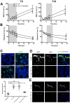
Effect of compstatin treatment on blood cells. Time course of WBC (A) and platelet (B) counts in the blood of baboons treated with compstatin during the first (T0) and second (T + 5) stage of experimental sepsis. Data are presented as mean ± SEM; 2-tailed Student t test; *P < .05. (C) Immunostaining and quantitation of CD68 positive macrophages in the lung of healthy controls, septic baboons (E coli), and septic baboons treated with compstatin (CS) during the first (E coli + CS T0) or the second (E coli + CS T + 5) stage. Scatter-plot data are shown as mean ± SEM; 1-way ANOVA with Dunnett multicomparison test. ***P < .001 compared with E coli group. (D-E) Immunostaining for C5b9 neo-antigen showing TCC deposition on platelets (D) or vascular endothelium (E) in the lung of septic baboons without (E coli) vs with compstatin (CS) treatment during second stage (E coli + CS T + 5). Note the absence of TCC on gpIIIa-positive platelets in compstatin-treated animals. Magnification bars for all images, 50μm.
Figure 4
Figure 4
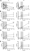
Time course of hemostatic parameters. (A, fibrinogen; B, FDP; C, APTT) and plasma levels of soluble P-selectin (D) and soluble thrombomodulin (E) in baboons treated with compstatin during the first (T0) and second (T + 5) stages of experimental sepsis. Data are presented as mean ± SEM; 2-tailed Student t test. *P < .05.
Figure 5
Figure 5
Localization and quantitative analysis of hemostatic proteins in the lung. (A) Micrograph panel showing (columns) immunostaining for PAI-1, tissue factor (TF), TFPI and thrombomodulin (TM) in (rows) healthy controls, septic baboons (E coli), and septic baboons treated with compstatin (CS) during the first (E coli + CS T0) or the second (E coli + CS T + 5) stage. The colors of the each antigen and nuclear counterstaining are shown on the top row. ct indicates convolulted tubules; and g, glomerulus. Magnification bars, 50 μm. (B) Scatter-plot representations of MFI of images collected for the above-mentioned proteins and experimental conditions. PAI-1: whole-field MFI of at least 15 images; all other immunostainings: 20 ROI (alveolar capillaries) in 15 images (200 spots). (C) Histogram representation of mRNA expression for PAI-1, TF, TFPI, and TM. Values indicate the mean ± SEM of fold over β-actin housekeeping gene. In panels B and C, data are presented as mean ± SEM. One-way ANOVA with Dunnett multicomparison test. *P < .05; **P < .001; ***P < .001 compared with E coli group.
Figure 6
Figure 6
Time-course of organ function and biochemical markers in the blood of baboons treated with compstatin during the first (T0) and second (T + 5) stage of experimental sepsis. (A) Mean systemic arterial pressure (MSAP); (B) creatinine; (C) lactate dehydrogenase (LDH); (D) alanine aminotransferase (ALT); (E) aspartate transaminase (AST). Data are presented as mean ± SEM; 2-tailed Student t test. *P < .05; **P < .01; ***P < .001.
Figure 7
Figure 7
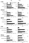
Comparison of the histopathologic changes in organs from septic animals with or without compstatin treatment during the first (T0) or second (T + 5) stage. The tissues were collected after euthanasia at T + 24 hours. Evaluations of the parameters were performed in a blinded fashion and graded on a scale from 0 to 4, with 0 being normal and 4 being severe. The histopathologic changes of the tissues collected from the 2 compstatin-treated groups are significantly less severe than those of the E coli challenge group. P < .001, with the exception of the lung congestion. Data are presented as mean ± SEM; 2-tailed Student t test. *P < .05; **P < .01; ***P < .001.

References

    1. Ward PA, Gao H. Sepsis, Complement, and the Dysregulated Inflammatory Response. J Cell Mol Med. 2009 - PMC - PubMed
    1. Angus DC, Linde-Zwirble WT, Lidicker J, Clermont G, Carcillo J, Pinsky MR. Epidemiology of severe sepsis in the United States: analysis of incidence, outcome, and associated costs of care. Crit Care Med. 2001;29(7):1303–1310. - PubMed
    1. Taylor FB., Jr Staging of the pathophysiologic responses of the primate microvasculature to Escherichia coli and endotoxin: examination of the elements of the compensated response and their links to the corresponding uncompensated lethal variants. Crit Care Med. 2001;29(7 Suppl):S78–89. - PubMed
    1. Markiewski MM, DeAngelis RA, Lambris JD. Complexity of complement activation in sepsis. J Cell Mol Med. 2008;12(6A):2245–2254. - PMC - PubMed
    1. Markiewski MM, Lambris JD. The role of complement in inflammatory diseases from behind the scenes into the spotlight. Am J Pathol. 2007;171(3):715–727. - PMC - PubMed

Publication types

MeSH terms