. 2010 Sep;30(9):1695-702.
doi: 10.1161/ATVBAHA.110.203034. Epub 2010 May 13.

Role of delta-like-4/Notch in the formation and wiring of the lymphatic network in zebrafish

Affiliations

Role of delta-like-4/Notch in the formation and wiring of the lymphatic network in zebrafish

Ilse Geudens et al. Arterioscler Thromb Vasc Biol. 2010 Sep.

Abstract

Objective: To study whether Notch signaling, which regulates cell fate decisions and vessel morphogenesis, controls lymphatic development.

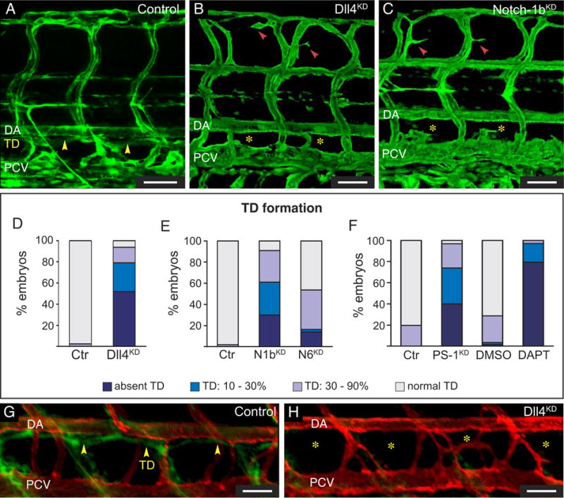
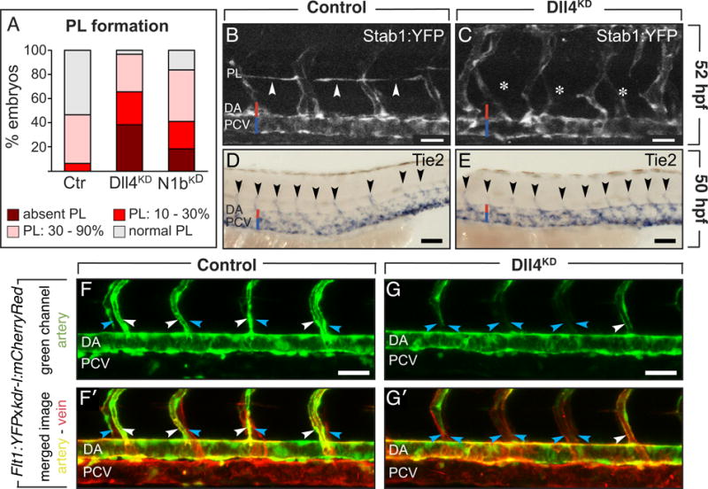
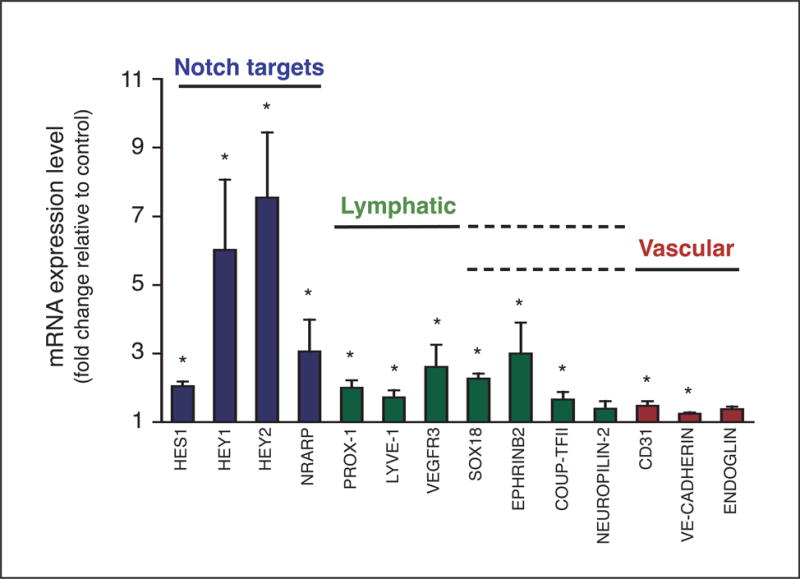
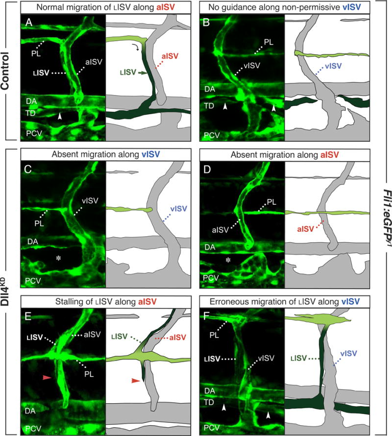
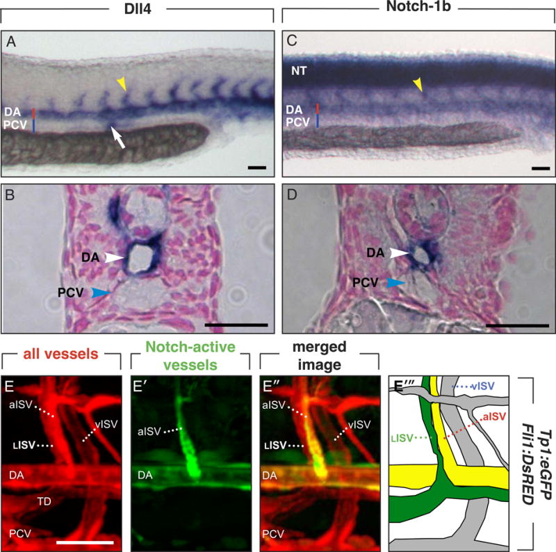
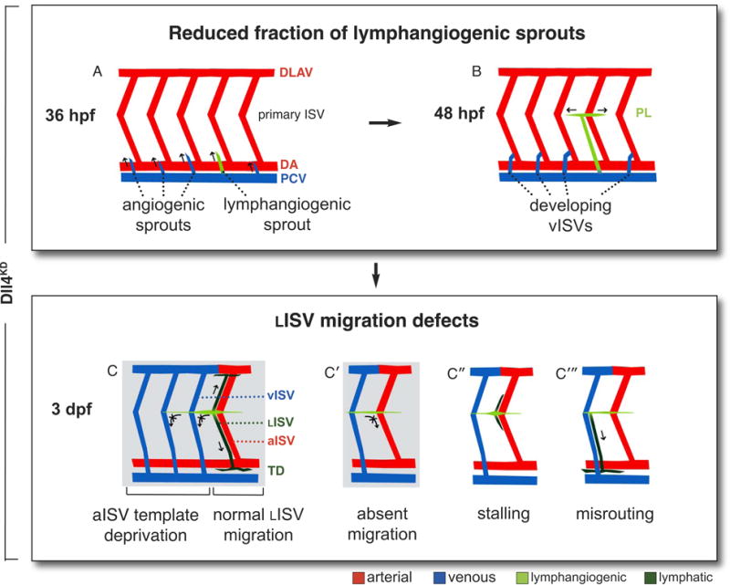
Methods and results: In zebrafish embryos, sprouts from the axial vein have lymphangiogenic potential because they give rise to the first lymphatics. Knockdown of delta-like-4 (Dll4) or its receptors Notch-1b or Notch-6 in zebrafish impaired lymphangiogenesis. Dll4/Notch silencing reduced the number of sprouts producing the string of parchordal lymphangioblasts; instead, sprouts connecting to the intersomitic vessels were formed. At a later phase, Notch silencing impaired navigation of lymphatic intersomitic vessels along their arterial templates.

Conclusions: These studies imply critical roles for Notch signaling in the formation and wiring of the lymphatic network.


Conflict of interest statement

Disclosure: The authors declare no competing financial interests.

Figures

Figure 1
Figure 1. Role of Notch in TD formation
A–C, Confocal images of GFP+ vessels in Fli1:eGFPy1 embryos. Normal TD in control (yellow arrowheads; A), and absent TD in Dll4KD (B) and Notch-1bKD (C) embryos. Yellow asterisks: TD absence; red arrowheads: minimal hyperbranching of ISVs. D–F, Percentage of affected embryos in control (N=122 in D; 185 in E; 87 in F) or Dll4KD embryos (N=80; 10ng Dll4SPL; P<0.001; D); Notch-1bKD (N1bKD) embryos (N=84; 15ng Notch-1bSPL; P<0.001; E), Notch-6KD (N6KD) embryos (N=63; 15ng Notch-6SPL; P<0.001; E), PS-1KD embryos (N=65; 2.5ng PS-1ATG1; P<0.001; F), or embryos treated with DMSO (N=171; F) or DAPT (N=34; 25 μM; P<0.001; F). G,H, Lymphangiography in 7-dpf kdr-l:mCherryRed embryos revealed normal uptake and drainage of a green dye by the TD in the control (yellow arrowheads; G), but not in the Dll4KD embryo (yellow asterisks; H). Bars: 50 μm.
Figure 2
Figure 2. Silencing of Notch blocks PL and lymphangiogenic sprout formation
A, Percentage of 52-hpf embryos with affected PL in control (N=73), Dll4KD embryos (N=55; 10ng Dll4SPL; P<0.001) or Notch-1bKD (N1bKD) embryos (N=49; 20ng Notch-1bATG; P<0.001). B,C, Confocal images of 52-hpf Stab1:YFP embryos, showing normal PL in controls (arrowheads; B), but absence in Dll4KD embryos (asterisks; C). D,E, Whole-mount in situ Tie2 staining at 50 hpf, revealing normal numbers of secondary sprouts (arrowheads) in control (D) and Dll4KD (E) embryos. F,G, Confocal images of vessels in Flt1:YFPxkdr-l:mCherryRed embryos: kdr-l:mCherryRed marks venous and arterial vessels red (red channel not shown), Flt1:YFP labels arterial vessels green (F,G), and merged images show arterial vessels yellow and venous vessels red (F′,G′). Lateral views with left and right side ISVs partially superimposed. Imaging was at 54 hpf, when secondary angiogenic sprouts had already connected to primary ISVs, which were changing arterial to venous identity in a ventral-to-dorsal pattern. In controls (F,F′), half of the aISVs became connected by angiogenic sprouts from the PCV and acquired a venous identity, thereby losing their green arterial signal (blue arrows; F) and becoming red only (blue arrows; F′), while the other half of the ISVs remained connected to the DA and were green (white arrows; F) or yellow in the merged image (white arrows; F′). By contrast, in the Dll4KD embryo (G,G′), most ISVs lost their green arterial marker (blue arrows; G; note the single white arrow), and became marked in red only (blue arrows in G′). White arrow in G,G′ denotes a residual aISV retaining its green (G) or yellow (G′) label. Bars: 50μm (B,C,F,G); 100μm (D,E).
Figure 3
Figure 3. Notch activation by Dll4 promotes lymphatic characteristics in vitro
RT-PCR of HUVECs, co-cultured with COS cells expressing hDll4 (COSDll4) or control GFP (COSCTR), confirming upregulation of Notch targets (HES1, HEY1, HEY2, NRARP; blue bars) and revealing enhanced lymphatic marker expression (PROX-1, LYVE-1, VEGFR3, SOX18, EPHRINB2, green bars), while vascular genes (CD31, VE-CADHERIN; ENDOGLIN; red bars) were only minimally affected. COUP-TFII was also upregulated. NEUROPILIN-2 was not affected. Lymphatic/arterial and lymphatic/venous genes are marked by the overlapping dashed lines. Results are fold change in HUVEC/COSDll4 co-culture versus HUVEC/COSCTR. Mean±SEM; N=3–11; *, P<0.05.
Figure 4
Figure 4. Incomplete silencing of Notch perturbs lymphatic navigation
Confocal images with accompanying schematic redrawing of the navigation routes of LISVs along aISVs or vISVs in 4-dpf control (A,B) and Dll4KD (C–F) Fli1:eGFPy1 embryos. Permanent lymphatic structures (LISV, TD) are dark green; transient lymphangiogenic structures (PL) are light green. A,B, In control embryos, LISV-PLs navigate alongside aISVs and establish a continuous TD (arrowhead). Note how LISVs “creep” over their aISV guidance templates (A). LISV-PLs never navigate along vISVs in control embryos (B). C–F, Navigation defects in Dll4KD embryos. C, In a large fraction of morphant somites, LISV-PLs lack migration templates because fewer aISVs develop. As LISV-PLs do not normally migrate along vISVs, no TD was formed in these somites (asterisks). D, In other morphant somites, LISV-PLs bypassed the point of turning at the aISVs, and failed to switch to radial migration. E, In a small fraction of somites, LISV-PLs accomplished to make the turn and switched to radial migration, but then stalled (red arrowhead denotes the arrested tip of a navigating LISV). F, In most Dll4KD embryos, vISVs were not permissive to guide LISV-PLs, but, occasionally, LISV-PLs erroneously navigated alongside a vISV.
Figure 5
Figure 5. Expression of Dll4./.Notch-1b
A–D, Whole-mount embryos at 30 hpf, when lymphangiogenic sprouting occurs, stained for Dll4 (A,B) or Notch-1b (C,D); panels B,D show cross-sections of the respective embryos. Primary ISVs are indicated by yellow arrowheads (A,C). A,B, Dll4 expression was detected in the DA and primary aISVs, pronephric duct (white arrow; A). C,D, Notch-1b is strongly expressed in the neural tube (NT), DA and primary aISVs. E, Confocal images of Tp1bglob:eGFPxFli1:DsRed embryos, in which Fli1:DsRed marks blood and lymph vessels in red (E) and Tp1bglob:eGFP labels cells with activated canonical Notch activity in green (E′). The merged image shows arterial vessels (DA; aISV) with active Notch in yellow (green-red), while the LISV are only red (E”). In the schematic representation, the lymphatic structures are indicated in green, Notch-activated vessels in yellow, and other vessels in grey (E‴). Representative images of arterial activation of Notch in a 6-dpf embryo are shown (for technical reasons), but similar data were obtained at 60 hpf. Bars: 50 μm.
Figure 6
Figure 6. Schematic model of Notch in lymphatic development
Scheme, illustrating the different lymphatic defects in Dll4KD embryos (normal lymphatic development, Supplemental Figure I). Permanent lymphatic structures (LISV, TD) are dark green; transient lymphangiogenic structures (lymphangiogenic secondary sprouts; parachordal lymphangioblasts) are light green. A,B, REDUCED FRACTION OF LYMPHANGIOGENIC SPROUTS, resulting in underdevelopment or absence of the PL string, with accompanying overrepresentation of angiogenic secondary sprouts. C–C‴, LISV MIGRATION DEFECTS: C, As a result of vISV overrepresentation, LISV-PLs are deprived of their normal aISV guidance template. C′–C‴, LISV formation is further impaired by additional navigation defects, most frequently because LISV-PLs cells bypass their turning point and never initiate ventral radial migration (C′), or occasionally make the turn but then stall (C”). More rarely, navigating LISV-PLs become misrouted along vISVs (C‴). The most frequent defects are boxed in grey.

Comment in

References

    1. Tammela T, Alitalo K. Lymphangiogenesis: Molecular mechanisms and future promise. Cell. 2010;140:460–476. - PubMed
    1. Francois M, Koopman P, Beltrame M. SoxF genes: Key players in the development of the cardio-vascular system. Int J Biochem Cell Biol. 2010;42:445–448. - PubMed
    1. Gale NW, Thurston G, Hackett SF, Renard R, Wang Q, McClain J, Martin C, Witte C, Witte MH, Jackson D, Suri C, Campochiaro PA, Wiegand SJ, Yancopoulos GD. Angiopoietin-2 is required for postnatal angiogenesis and lymphatic patterning, and only the latter role is rescued by Angiopoietin-1. Dev Cell. 2002;3:411–423. - PubMed
    1. De Smet F, Segura I, De Bock K, Hohensinner PJ, Carmeliet P. Mechanisms of vessel branching: filopodia on endothelial tip cells lead the way. Arterioscler Thromb Vasc Biol. 2009;29:639–649. - PubMed
    1. Tammela T, Saaristo A, Lohela M, Morisada T, Tornberg J, Norrmen C, Oike Y, Pajusola K, Thurston G, Suda T, Yla-Herttuala S, Alitalo K. Angiopoietin-1 promotes lymphatic sprouting and hyperplasia. Blood. 2005;105:4642–4648. - PubMed

Publication types

MeSH terms