. 2010 Aug;9(8):1666-77.
doi: 10.1074/mcp.R000002-MCP201. Epub 2010 May 13.

Toward an integrated structural model of the 26S proteasome

Affiliations

Toward an integrated structural model of the 26S proteasome

Friedrich Förster et al. Mol Cell Proteomics. 2010 Aug.

Abstract

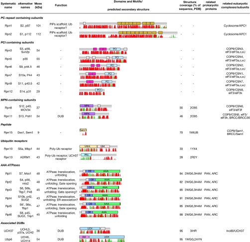
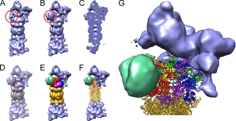
The 26S proteasome is the end point of the ubiquitin-proteasome pathway and degrades ubiquitylated substrates. It is composed of the 20S core particle (CP), where degradation occurs, and the 19S regulatory particle (RP), which ensures substrate specificity of degradation. Whereas the CP is resolved to atomic resolution, the architecture of the RP is largely unknown. We provide a comprehensive analysis of the current structural knowledge on the RP, including structures of the RP subunits, physical protein-protein interactions, and cryoelectron microscopy data. These data allowed us to compute an atomic model for the CP-AAA-ATPase subcomplex. In addition to this atomic model, further subunits can be mapped approximately, which lets us hypothesize on the substrate path during its degradation.


Figures

Fig. 1.
Fig. 1.
Overview of RP subunits. PC repeat (orange), coiled coils (white, stripes), TPR-like repeats (magenta), PCI module, MPN domain, Sem1 fold, von Willebrand domain (VWA), UIM, PRU, OB fold, AAA-ATPase domain, UCH domain, and ubiquitin-like domain (Ubl) are shown. Secondary structures (red, α-helical; green, β-strand) were predicted using the MPI bioinformatics toolkit (109) as a front end to PSIPRED (110). Putative coiled coils with probabilities >0.5 according to PCOILS (111) are indicated. TPR motifs were assigned using TPRpred (80). ER, endoplasmic reticulum; PIP, proteasome interacting protein.
Fig. 2.
Fig. 2.
Structures or probable folds of RP subunits. A, AAA-ATPases. Crystal structures of PAN-N (orange) and the PAN AAA fold (red-orange) were determined separately (Protein Data Bank codes 2WG6 and 3H4M). B, Rpn1 and Rpn2. The PC repeats of Rpn1 and Rpn2 probably adopt a solenoid fold similar to protein phosphatase PP2A (residues 282–584 of Protein Data Bank code 1B3U; sequence identity, 15%). C, PCI-module containing subunits. The PCI module of the COP9 subunit CSN7 (Protein Data Bank code 3CHM) consists of a C-terminal winged helix domain (light blue) and a helical bundle (blue). In CSN7, as probably also in most proteasomal PCI subunits, the PCI module is preceded by one or more bihelical domains (light red). D, Rpn8 and Rpn11. The MPN domain of Rpn8 (Protein Data Bank code 2O95) is shown. E, ubiquitin receptors. The Rpn10 UIM (blue) consists of three helices that lack a defined tertiary structure (Protein Data Bank code 1YX5). Ubiquitin (red) binds to the UIM helices. In contrast, ubiquitin binds to loops of the globular PRU domain of Rpn13 (blue). F, associated DUBs. UCH37 exhibits a globular UCH fold and a C-terminal domain, which contains a coiled coil motif. In Ubp6, a UCH fold (Protein Data Bank code 2AYN) is preceded by an N-terminal Ub-like domain (Protein Data Bank code 1WGG) both of which have been solved separately.
Fig. 3.
Fig. 3.
Cryo-EM density map of D. melanogaster 26S proteasome. A, image classification revealed that ∼50% of all analyzed particles lacked an additional density (red circle). B, the remaining 50% possessed an additional density (red circle). C, side cut of density depicted in B. D, atomic models of the CP (gold) and the AAA-ATPases (Rpt1/Rpt2/Rpt6/Rpt3/Rpt4/Rpt5, red/orange/yellow/green/blue/purple) fitted into map B. E, segmentation of B according to the fitted atomic models. F, hybrid representation of the 26S proteasome by atomic models and the cryo-EM density where interpretation on the atomic level is not possible yet. G, enlarged view of F.
Fig. 4.
Fig. 4.
RP topology based on publicly available protein-protein interaction data (Table I). The protein-protein interactions were determined by two-hybrid assays (red), in vivo pulldown experiments (orange), chemical cross-linking (dashed black), in vitro binding (magenta), and co-expression (green). Dotted lines indicate interactions involving more than two proteins.
Fig. 5.
Fig. 5.
Schematic representation of protein degradation through the 26S proteasome. Ubiquitylated substrates first bind to the Ub receptors Rpn10 and Rpn13. As the substrates proceed, they get deubiquitylated by Rpn11, then unfolded in the upper ring of the AAA-ATPase hexamer, and translocated to the CP by the lower AAA-ATPase domains. The AAA-ATPase subunits Rpt2 and Rpt5 open the gate to the CP where substrates get cleaved by CP subunits β1, β2, and β5 located in the CP inner cavity (8).

Similar articles

Cited by

References

    1. Murata S., Yashiroda H., Tanaka K. (2009) Molecular mechanisms of proteasome assembly. Nat. Rev. Mol. Cell Biol. 10, 104–115 - PubMed
    1. Glickman M. H., Ciechanover A. (2002) The ubiquitin-proteasome proteolytic pathway: destruction for the sake of construction. Physiol. Rev. 82, 373–428 - PubMed
    1. Voges D., Zwickl P., Baumeister W. (1999) The 26S proteasome: a molecular machine designed for controlled proteolysis. Annu. Rev. Biochem. 68, 1015–1068 - PubMed
    1. Jentsch S., Haendler B. (2009) The Ubiquitin System in Health and Disease, Springer, Heidelberg, Germany - PubMed
    1. Pühler G., Weinkauf S., Bachmann L., Müller S., Engel A., Hegerl R., Baumeister W. (1992) Subunit stoichiometry and three-dimensional arrangement in proteasomes from Thermoplasma acidophilum. EMBO J. 11, 1607–1616 - PMC - PubMed

Publication types

MeSH terms

Substances

LinkOut - more resources