. 2010 Jun;69(6):606-22.
doi: 10.1097/NEN.0b013e3181e00579.

Differential distribution of erbB receptors in human glioblastoma multiforme: expression of erbB3 in CD133-positive putative cancer stem cells

Affiliations

Differential distribution of erbB receptors in human glioblastoma multiforme: expression of erbB3 in CD133-positive putative cancer stem cells

Véronique Duhem-Tonnelle et al. J Neuropathol Exp Neurol. 2010 Jun.

Erratum in

  • J Neuropathol Exp Neurol. 2010 Nov;69(11):1176

Abstract

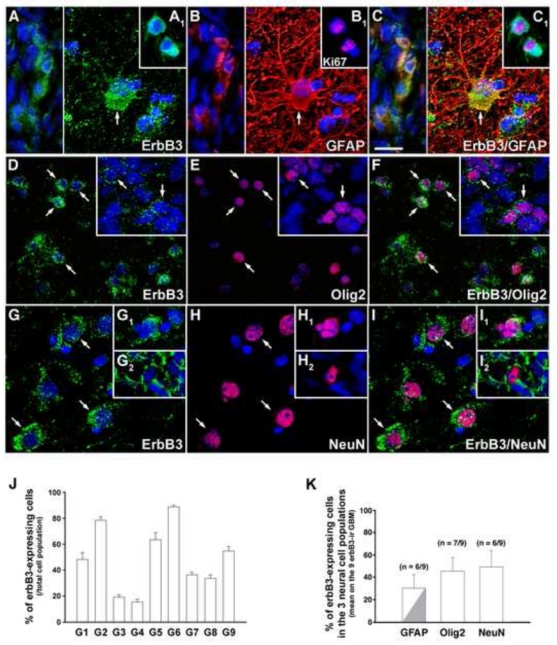
Glioblastomas are the most common primary central nervous system tumors in adults, and they remain resistant to current treatments. erbB1 signaling is frequently altered in glioblastomas, suggesting thaterbB receptor family members may represent targets for molecular therapy. We performed a comprehensive analysis of erbB receptor and ligand expression profiles in a panel of 9 glioblastomas andcompared them to nonneoplastic cerebral tissue containing neocortex and adjacent white matter. Quantitative reverse transcription-polymerase chain reaction and Western blot analysis showed that erbB1signaling and erbB2 receptors exhibited highly variable deregulation profiles in the tumors, with patterns ranging from underexpression to overexpression; in contrast, erbB3 and erbB4 were downregulated. We next performed immunohistochemistry to determinethe distribution patterns of erbB receptors among the main neuralcell types in the tumors with special reference to the putative tumor stem cell population. Results revealed intertumoral and intratumoral heterogeneity in all 4 erbB expression profiles, but each receptor exhibited a distinct distribution pattern among glial fibrillary acidic protein-, Olig2-, NeuN-, and CD133-positive populations. Although erbB1 immunoreactivity was detected in only small subsets of CD133-positive putative tumor stem cells, erbB3 immunoreactivity was prominent in this population, suggesting that erbB3 may represent a new potential therapeutic target.


Figures

Figure 1
Figure 1
Messenger RNA (mRNA) expression of erbB signaling genes in glioblastomas as compared to control cortices. Real-time quantitative RT-PCR analysis of the four ERBB genes and their eleven ligands (EGF, TGFα, AREG, EPGN, HB-EGF, BTC, EREG, NRG1, NRG2, NRG3 and NRG4) in the nine glioblastoma samples (G1 to G9). Messenger RNA levels of three zinc-dependent matrix metalloproteinases that are involved in ectodomain shedding of erbB ligands (MMP2, MMP9 and ADAM17) and of the KI67 cellular proliferation marker were also evaluated. Results are expressed as the fold increase in gene expression level in the nine glioblastoma samples relative to the mean respective expression level in the four non-neoplastic cortical tissues. Fold expressions were scored as described in the colorimetric scale and the materials and methods section. Note that, although ERBB1 receptor and ligands and ERBB2 receptor mRNA exhibit highly heterogeneous deregulation profiles between tumors, most NRG signaling genes globally show underexpression in our panel of glioblastomas as compared to control cortices. NE, non-expressed.
Figure 2
Figure 2
Protein expression of erbB receptors in glioblastoma tissues and cell lines and in control samples. (A) Western blot analysis of erbB1-4 receptors expression in control non-neoplastic samples (primary cultures of fetal cortical (Ctx) and hypothalamic (Hyp) astrocytes, fetal brain, and the four cerebral cortex samples C1 to C4) and in neoplastic samples (the nine glioblastomas G1 to G9, and three human glioblastoma cell lines (hGBM cell lines)). Eighty-five micrograms of proteins were loaded into each well. Three primary cultures of fetal cortical, hypothalamic astrocytes and three fetal brains were analyzed. One representative sample of each is shown (B–E) Densitometric analysis of erbB1 (B), erbB2 (C), erbB3 (D) and erbB4 (E) protein expression in the four control cortical tissues (Cortex) and the nine glioblastoma samples. X axis, sample number. Results are expressed as the fold increase in erbB protein expression for each glioblastoma sample (black columns) relative to the mean respective erbB protein expression in the four non-neoplastic cortical tissues (white columns). Horizontal solid line, mean erbB protein expression in the four control cortical samples normalized to 1. Note that erbB1 and erbB2 receptors exhibit highly discrepant expression profiles among tumoral samples, while erbB3 and erbB4 protein contents are consistently down-regulated in our glioblastoma panel. (F–I) For each erbB receptor, the fold mRNA expression values were compared to the respective fold protein expression values across the glioblastoma cohort using a linear regression test. Note the overall correlation between mRNA and protein levels of erbB receptors in tumors relative to control cortices.
Figure 3
Figure 3
In the human adult cerebral cortex, erbB3 expression is detected in neurons, oligodendrocytes and astrocytes, whereas erbB4 expression is restricted to a sub-population of neurons. (A–I) Double immunofluorescent labeling of erbB3 (green) with NeuN (red), Olig2 (red) or GFAP (red) shows an expression of erbB3 in neurons, oligodendrocytes and astrocytes, respectively (arrows). The asterisks point to an Olig2-positive oligodendrocyte devoid of erbB3 immunoreactivity in the white matter (D–F) and a GFAP-positive astrocyte lacking erbB3 immunoreactivity in the grey matter (G–I). (J–L) Double immunofluorescent labeling of erbB4 (green) and NeuN (red) reveals that erbB4 is expressed in a subset of neurons (arrows) scattered among erbB4-negative neurons (asterisk) in the cortex. Note that NeuN is detected both in the nucleus and the soma cytoplasm of neurons (B, K), as described elsewhere (67). Nuclei were counter-stained with Hoechst (blue). Scale bar = 20 μm.
Figure 4
Figure 4
ErbB1 expression is detected in all glioblastoma samples with highly variable immunoreactivity profiles but is preferentially expressed in the GFAP- and Olig2-positive cellular contingents. (A–F) Double immunofluorescent labeling of erbB1 (green) with GFAP (red) or Olig2 (red) reveals the presence of erbB1 in a fraction of the GFAP-positive cell population (A–C, arrows) and of the Olig2-positive cell population (D–F, arrows). Nuclei were counter-stained with Hoechst (blue). Scale bar = 20 μm. (G) Quantification of erbB1 expression in the nine glioblastoma samples. Results are expressed as the % number of erbB1-immunoreactive cells over the total number of Hoechst-positive nuclei. Vertical bars represent SEM. (H) Quantification of erbB1 expression in GFAP-, Olig2- and NeuN-positive cell populations (see materials and methods). Bar graph represents the mean % of erbB1-immunoreactive cells on the nine erbB1-immunoreactive (ir) glioblastomas (GBM) in each cell type. Vertical bars represent SEM.
Figure 5
Figure 5
ErbB2 immunoreactivity is detected in all glioblastoma samples with heterogeneous tissue and cellular expression profiles among samples. (A–I) Double immunofluorescent labeling of erbB2 (green) with GFAP (red), Olig2 (red) or NeuN (red) reveals that erbB2 is expressed in GFAP- (A–C, arrows), Olig2- (D–F, arrows) and NeuN-positive cells (G–I, arrows). Note that erbB2 is detected in NeuN-immunoreactive cells that exhibit a big round nucleus reminiscent of those seen in the normal cortex (G–I, inset) or in NeuN-positive nuclei of small size and heterogeneous shapes (G–I, main panel). Nuclei were counter-stained with Hoechst (blue). Scale bar = 20 μm. (J) Quantification of erbB2 expression in the nine glioblastoma samples. Results are expressed as the % number of erbB2-immunoreactive cells over the total number of Hoechst-positive nuclei. Vertical bars represent SEM. (K) Quantification of erbB2 expression in GFAP-, Olig2- and NeuN-positive cell populations (see materials and methods). Bar graph represents the mean % of erbB2-immunoreactive cells on the nine erbB2-immunoreactive (ir) glioblastomas (GBM) in each cell type. Vertical bars represent SEM.
Figure 6
Figure 6
In the glioblastomas, erbB3 is expressed in significant fractions of the GFAP-, Olig2- and NeuN-positive cell populations. (A–I) Double immunofluorescent labeling of erbB3 (green) with GFAP (red), Olig2 (red) or NeuN (red) reveals the presence of erbB3 in GFAP- (A–C, arrow and left panel), Olig2- (D–F, arrows) and NeuN-positive cells (G–I, arrows and insets). (A1–C1) Double immunofluorescent labeling of erbB3 (green) with the proliferation marker Ki67 (red). Note that erbB3 is detected in Olig2- and NeuN-positive cells of various sizes and shapes and is expressed in GFAP-positive cells exhibiting small and irregular morphologies (A–C, left) or an hypertrophic shape reminiscent of reactive astrocytes (A–C, arrow). In tumors, all Ki67-positive cells were also immunoreactive for erbB3 (A1–C1). Nuclei were counter-stained with Hoechst (blue). Scale bar = 20 μm. (J) Quantification of erbB3 expression in the nine glioblastoma samples. Results are expressed as the % number of erbB3-immunoreactive cells over the total number of Hoechst-positive nuclei. Vertical bars represent SEM. (K) Quantification of erbB3 expression in GFAP-, Olig2- and NeuN-positive cell populations (see materials and methods). Bar graph represents the mean % of erbB3-immunoreactive cells on the nine erbB3-immunoreactive (ir) glioblastomas (GBM) in each cell type. A fraction of the erbB3/GFAP co-expressing cell population corresponded to hypertrophic reactive astrocytes (grey). Vertical bars represent SEM.
Figure 7
Figure 7
ErbB4 immunoreactivity is preferentially distributed in neuronal-like elements in the glioblastomas and is also occasionally detected in hypertrophic astrocytes. (A–F) Double immunofluorescent labeling of erbB4 (green) with GFAP (red) or NeuN (red). ErbB4 immunoreactivity mainly appeared as a bright punctuated staining of fine cell processes (A–C, arrowheads). Rare erbB4/NeuN co-expressing cells were detected in some tumors (D–F, inset). An atypical immunostaining profile consisting of numerous erbB4-weakly immunoreactive cell processes deprived of the usual punctuated aspect (D–F, main panel, arrows) was seen in a glioblastoma in association with abnormal NeuN-positive nuclei. ErbB4 immunoreactivity was occasionally detected in GFAP-positive hypertrophic astrocytes (A–C, arrows). Note that the erbB4 immunolabeling did not exhibit a punctuate aspect in GFAP-positive soma and cell processes. Nuclei were counter-stained with Hoechst (blue). Scale bar = 20 μm. (G) Quantification of erbB4 expression in GFAP-, Olig2- and NeuN-positive cell populations (see materials and methods). Bar graph represents the mean % of erbB4-immunoreactive cells on the eight erbB4-immunoreactive (ir) glioblastomas (GBM) in each cell type. Note that although all erbB4-expressing tumors contained erbB4-immunoreactive cell processes, only 3/8 exhibited erbB4/NeuN co-expressing soma. ErbB4/GFAP-positive elements only consisted of hypertrophic astrocytes (grey). Vertical bars represent SEM.
Figure 8
Figure 8
ErbB1 and erbB3 receptors are expressed in subsets of CD133-immunoreactive cells. (A–F) Double immunofluorescent labeling of erbB1 (A, C, green) and erbB3 (D, F, green) with CD133 (red) shows the presence of erbB1 (A–C, arrows) and erbB3 (D–F, arrows) in CD133-positive cells. Some CD133-positive cells were occasionally seen at the vicinity of blood vessels (A–C, asterisk). Nuclei were counter-stained with Hoechst (blue). Scale bar = 20 μm. (G) Quantification of CD133 expression in the nine glioblastoma samples. Results are expressed as the % number of CD133-immunoreactive cells over the total number of Hoechst-positive nuclei in CD133-positive tumoral areas. Vertical bars represent SEM. (H) Quantification of erbB1, erbB3 and erbB4 expression in the CD133-positive cell populations (see materials and methods). Bar graph represents the mean % of erbB-immunoreactive cells in the CD133-positive cell population on the seven CD133-immunoreactive (ir) glioblastomas (GBM). Vertical bars represent SEM.

References

    1. Wen PY, Kesari S. Malignant gliomas in adults. N Engl J Med. 2008;359:492–507. - PubMed
    1. Nicholas MK, Lukas RV, Jafri NF, et al. Epidermal growth factor receptor - mediated signal transduction in the development and therapy of gliomas. Clin Cancer Res. 2006;12:7261–7270. - PubMed
    1. Bernstein JJ, Anagnostopoulos AV, Hattwick EA, et al. Human-specific c-neu proto-oncogene protein overexpression in human malignant astrocytomas before and after xenografting. J Neurosurg. 1993;78:240–251. - PubMed
    1. Dietzmann K, von Bossanyi P. Coexpression of epidermal growth factor receptor protein and c-erbB-2 oncoprotein in human astrocytic tumors. An immunohistochemical study. Zentralbl Pathol. 1994;140:335–341. - PubMed
    1. Schlegel J, Stumm G, Brandle K, et al. Amplification and differential expression of members of the erbB-gene family in human glioblastoma. J Neurooncol. 1994;22:201–207. - PubMed

Publication types

MeSH terms