. 2008 Jun:20:19-37.
doi: 10.3767/003158508X302212. Epub 2008 Mar 21.

Multiple gene genealogies and phenotypic characters differentiate several novel species of Mycosphaerella and related anamorphs on banana

Affiliations

Multiple gene genealogies and phenotypic characters differentiate several novel species of Mycosphaerella and related anamorphs on banana

M Arzanlou et al. Persoonia. 2008 Jun.

Abstract

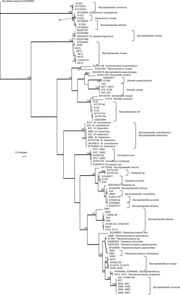
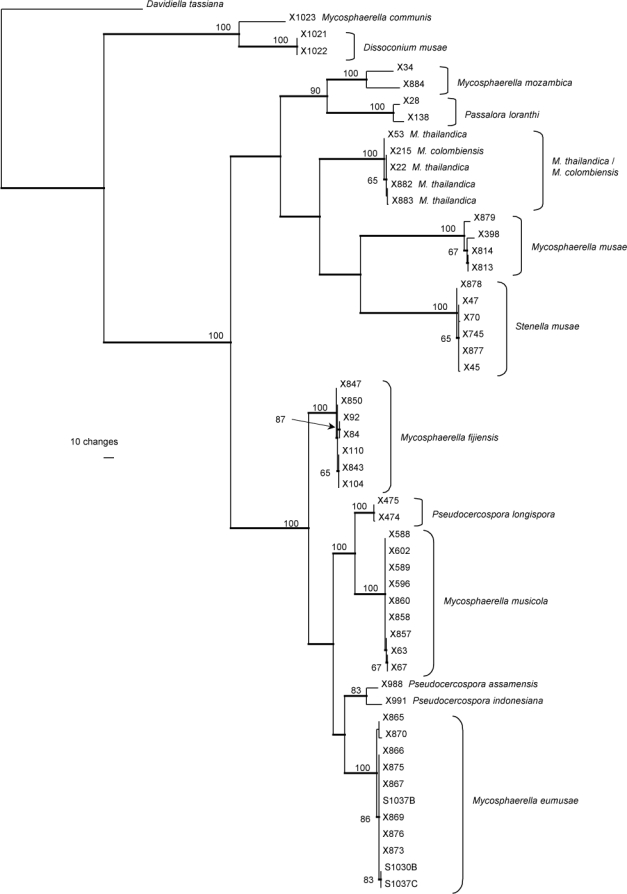
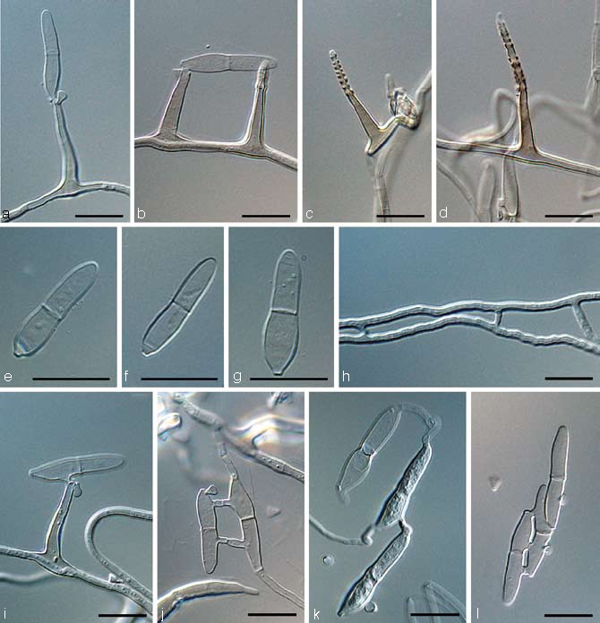
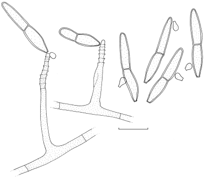
Three species of Mycosphaerella, namely M. eumusae, M. fijiensis, and M. musicola are involved in the Sigatoka disease complex of bananas. Besides these three primary pathogens, several additional species of Mycosphaerella or their anamorphs have been described from Musa. However, very little is known about these taxa, and for the majority of these species no culture or DNA is available for study. In the present study, we collected a global set of Mycosphaerella strains from banana, and compared them by means of morphology and a multi-gene nucleotide sequence data set. The phylogeny inferred from the ITS region and the combined data set containing partial gene sequences of the actin gene, the small subunit mitochondrial ribosomal DNA and the histone H3 gene revealed a rich diversity of Mycosphaerella species on Musa. Integration of morphological and molecular data sets confirmed more than 20 species of Mycosphaerella (incl. anamorphs) to occur on banana. This study reconfirmed the previously described presence of Cercospora apii, M. citri and M. thailandica, and also identified Mycosphaerella communis, M. lateralis and Passalora loranthi on this host. Moreover, eight new species identified from Musa are described, namely Dissoconium musae, Mycosphaerella mozambica, Pseudocercospora assamensis, P. indonesiana, P. longispora, Stenella musae, S. musicola, and S. queenslandica.

Keywords: Mycosphaerella; Sigatoka disease complex; phylogeny; taxonomy.


Figures

Fig. 1
Fig. 1
One of 11 780 equally most parsimonious trees obtained from a heuristic search with simple taxon additions of the ITS sequence alignment. The scale bar shows 10 changes, and bootstrap support values (65 % and higher) from 10 000 000 fast stepwise replicates are shown at the nodes. Thickened lines indicate the strict consensus branches. The tree was rooted to sequences of Davidiella tassiana strain CPC 11600 (GenBank accession number DQ289800). M. = Mycosphaerella and Ps. = Pseudocercospora.
Fig. 2
Fig. 2
One of eight equally most parsimonious trees obtained from a heuristic search with 100 random taxon additions of the combined (ITS, ACT, HIS, mtSSU) sequence alignment. The scale bar shows 10 changes, and bootstrap support values (65 % and higher) from 1000 replicates are shown at the nodes. Thickened lines indicate the strict consensus branches. The tree was rooted to sequences of Davidiella tassiana strain CPC 11600 (GenBank accession number DQ289800, DQ289867, EF679665, EU514455, respectively). M. = Mycosphaerella.
Fig. 3
Fig. 3
Dissoconium musae (CBS 122453). a–d. Conidiophores with sympodially proliferating conidiogenous cells, which produce primary and secondary conidia in pairs; e–g. primary conidia with truncate base; h–l. anastomoses between hyphae, primary and secondary conidia and primary conidia. — Scale bar = 10 μm.
Fig. 4
Fig. 4
Dissoconium musae (CBS 122453). — Scale bar = 10 μm.
Fig. 5
Fig. 5
Mycosphaerella mozambica (CBS 122464). a. Verruculose hyphae; b–e. unbranched or loosely branched conidiophores with sympodially proliferating conidiogenous cells; f–g. sympodially proliferating conidiogenous cells give rise to short conidium-bearing rachis; h. conidia with truncate base. — Scale bars = 10 μm.
Fig. 6
Fig. 6
Mycosphaerella mozambica (CBS 122464). a. Ascus with biseriate ascospores; b. ascospore germination pattern; c. conidiophores with sympodially proliferating conidiogenous cells, which give rise to short conidium-bearing rachis; d. conidia. — Scale bar = 10 μm.
Fig. 7
Fig. 7
Pseudocercospora assamensis (CBS 122467). a. Conidiophore with sympodial and percurrent growth of conidiogenous cell; b–c. conidia. — Scale bar = 10 μm.
Fig. 8
Fig. 8
Pseudocercospora assamensis (CBS 122467). — Scale bar = 10 μm.
Fig. 9
Fig. 9
Pseudocercospora indonesiana (CBS 122473). a–d. Conidia; e. intercalary conidiogenous cell. — Scale bar = 10 μm.
Fig. 10
Fig. 10
Pseudocercospora indonesiana (CBS 122473). — Scale bar = 10 μm.
Fig. 11
Fig. 11
Pseudocercospora longispora (CBS 122469). a–e. Conidia. — Scale bar = 10 μm.
Fig. 12
Fig. 12
Pseudocercospora longispora (CBS 122469). — Scale bar = 10 μm.
Fig. 13
Fig. 13
Stenella musae (CBS 122477). a–d. Conidiophores with sympodially proliferating conidiogenous cells; e–f. conidia. — Scale bar = 10 μm.
Fig. 14
Fig. 14
a. Stenella musae (CBS 122477); b. Stenella queenslandica (CBS 122475); c. Stenella musicola (CBS 122479). — Scale bar = 10 μm.
Fig. 15
Fig. 15
Ascospores of Mycosphaerella musae (IMI 91165). — Scale bar = 10 μm.
Fig. 16
Fig. 16
Stenella musicola (CBS 122479). a–e. Conidiophores with sympodially proliferating conidiogenous cells and darkened, thickened loci; f–g. hyphal anastomoses; h–i. conidia. — Scale bar = 10 μm.
Fig. 17
Fig. 17
Stenella queenslandica (CBS 122475). a. Conidiophore with terminal conidiogenous cell; b–d. conidia. — Scale bar = 10 μm.
Fig. 18
Fig. 18
a–c. Conidia in Pseudocercospora eumusae, P. fijiensis, and P. musae, respectively; d–f. ascospore germination pattern in M. eumusae, M. fijiensis and M. musicola, respectively. — Scale bar = 10 μm.
Fig. 19
Fig. 19
Pseudocercospora fijiensis. a–c. Conidiophores with sympodially proliferating conidiogenous cells; d–f. obclavate conidia with darkened hilum. — Scale bar = 10 μm.

References

    1. Aptroot A. 2006. Mycosphaerella and its anamorphs: 2. Conspectus of Mycosphaerella. CBS Biodiversity Series 5: 1 – 231 .
    1. Arzanlou M, Abeln ECA, Kema GHJ, Waalwijk C, Carlier J, Vries I de, Guzmán M, Crous PW. 2007a. Molecular diagnostics for the Sigatoka disease complex of banana. Phytopathology 97: 1112 – 1118 . - PubMed
    1. Arzanlou M, Groenewald JZ, Gams W, Braun U, Shin H-D, Crous PW. 2007b. Phylogenetic and morphotaxonomic revision of Ramichloridium and allied genera. Studies in Mycology 58: 57 – 93 . - PMC - PubMed
    1. Burgess TI, Barber PA, Sufaati S, Xu D, Hardy GEStJ, Dell B. 2007. Mycosphaerella spp. on Eucalyptus in Asia; new species; new hosts and new records. Fungal Diversity 24: 135 – 157 .
    1. Carbone I, Kohn LM. 1999. A method for designing primer sets for speciation studies in filamentous ascomycetes. Mycologia 91: 553 – 556 .

LinkOut - more resources