. 2010 Jun 10;114(22):7516-23.
doi: 10.1021/jp101415g.

A molecular view of the cholesterol condensing effect in DOPC lipid bilayers

Affiliations

A molecular view of the cholesterol condensing effect in DOPC lipid bilayers

Mohammad Alwarawrah et al. J Phys Chem B. .

Abstract

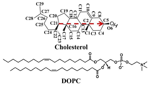
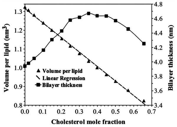
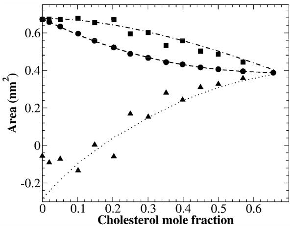
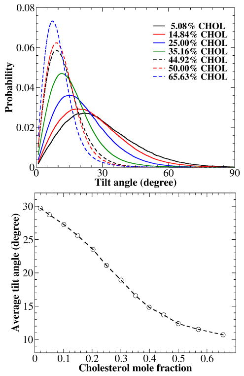
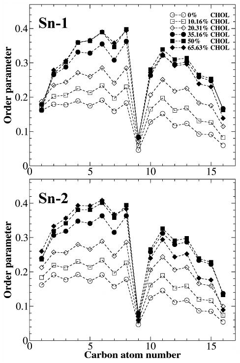
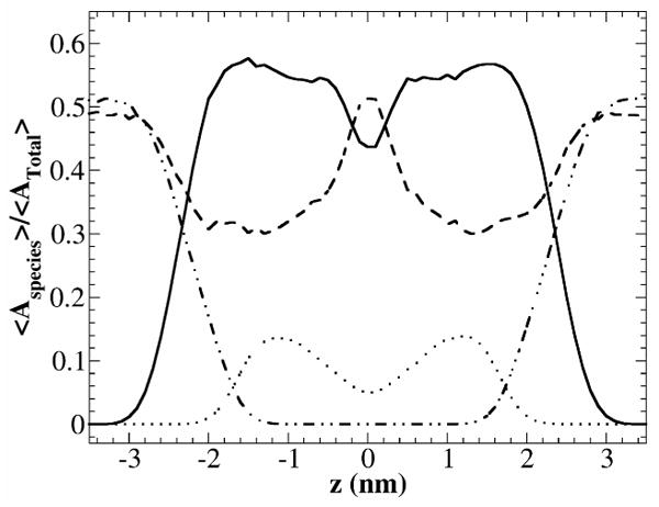
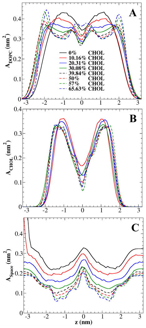
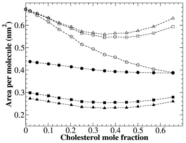
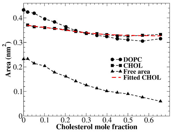
The condensing effect of cholesterol in dioleoylphosphatidylcholine (DOPC) lipid bilayers was systematically investigated via atomistic molecular dynamics (MD) simulation. Fourteen independent 200 ns simulations, spanning the entire range of cholesterol mole fraction (x(c)) in DOPC bilayers (i.e., from x(c) = 0 to 0.66), were performed at 323 K. The molecular areas occupied by DOPC and cholesterol at different distances from the bilayer center were analyzed using a slicing method based on the VDW radii of atoms. Curiously, while the average area per lipid and the cholesterol tilt angle, with respect to the bilayer normal, both show monotonic decreases as x(c) increases, the average bilayer height shows a significant decrease for x(c) > 0.35, following an initial increase. The calculated partial-specific areas of lipids clearly show the condensing effect of cholesterol. The VDW areal analysis showed that the condensing effect is limited only to the cholesterol sterol ring region, where the acyl chains of DOPC are severely compressed by cholesterol. As x(c) increases, the headgroups of DOPC gradually expand along the bilayer-aqueous interface to occupy more lateral area. Thus, it confirmed a key prediction of the umbrella model. At high cholesterol mole fractions, the calculated area per DOPC and area per cholesterol using some existing methods showed an inconsistent result: both increase while the overall area per lipid decreases. The inconsistency stems from the problematic assumption that cholesterol and DOPC have cylindrical shape and the same height. Our results showed that the total area of a PC/cholesterol bilayer is primarily determined by the molecular packing in the cholesterol sterol ring region. An alternative analysis of area per molecule within this region is proposed, which takes into account the cholesterol tilt angle and the practical incompressibility of cholesterol sterol rings. The new calculation shows that the majority of the area lost due to the cholesterol condensing effect is taken from PC molecules.


Figures

Figure 1
Figure 1
Chemical structures of cholesterol and DOPC. The vector connecting C21 and C5 atoms of cholesterol (red arrow) is used to represent the orientation of a cholesterol molecule, and the angle between this vector and the bilayer normal is defined as the cholesterol tilt angle.
Figure 2
Figure 2
Volume per lipid vpl(xc) and bilayer thickness h(xc) versus cholesterol mole fraction in DOPC bilayers. The linear fit to the volume per lipid data is: vpl(xc) =1.316-0.775xc.
Figure 3
Figure 3
Area per lipid and partial-specific areas of DOPC and cholesterol vs. cholesterol mole fraction. Circles: Average area per lipid; Square: DOPC partial-specific area; Triangles: Cholesterol partial-specific area.
Figure 4
Figure 4
(A) Tilt angle distributions between the bilayer normal and the vector connecting C21 and C5 atoms of cholesterol in DOPC bilayers at some selected cholesterol concentrations. (B) Average cholesterol tilt angle versus cholesterol mole fraction.
Figure 5
Figure 5
Acyl chain order parameter versus the carbon atom number in DOPC bilayers at some selected cholesterol concentrations.
Figure 6
Figure 6
The VDW areal fractions as functions of z for a DOPC bilayer containing 20.31 mol % of cholesterol. z is the distance from the bilayer center in the direction of bilayer normal. The areal fractions are normalized by total bilayer area. DOPC VDW areal fraction <ADOPC(z)>/<Atotal> (solid line); cholesterol VDW areal fraction <ACHOL(z)>/<Atotal> (dotted line); water areal fraction <Awater(z)>/<Atotal> (dot-dot-dash line); and free area fraction <Afree(z)>/<Atotal> (dashed line).
Figure 7
Figure 7
Snapshots of cross-sectional slices of a DOPC bilayer with 20.31 mol % of cholesterol at 200 ns. DOPC elements are colored in red; cholesterol in green, and water in blue. The remaining area is the free area. (A) z ≈ 0, i.e., at bilayer center; (B) z ≈ 1.2 nm, where many cholesterol sterol rings resided; (C) z ≈ 1.8 nm, where many cholesterol headgroups resided; (D) z ≈ 2.3 nm, where many DOPC headgroups resided.
Figure 8
Figure 8
The VDW area per DOPC (A), per cholesterol (B), and free space per lipid (C) versus location z at some selected cholesterol concentrations.
Figure 9
Figure 9
Area per DOPC and area per cholesterol calculated using different methods. Area per DOPC: by Method 1 (open squares) and by Method 2 (open triangles). Area per cholesterol: by Method 1 (filled squares) and by Method 2 (filled triangles). Area per cholesterol in the areal-determining planes (i.e., cholesterol sterol ring region): filled circles; Area per DOPC in the areal-determining planes: open circles.
Figure 10
Figure 10
The VDW areas of DOPC and cholesterol, and free area per lipid as functions of cholesterol mole fraction in the areal-determining planes. The red dash line is the fitting function Ao/cos(θ(xc)), where θ(xc) is the average cholesterol tilt angle plotted in Fig. 4B, and Ao is the fitting constant (0.3179±0.0026 nm2).

References

    1. Berkowitz ML. Biochim Biophys Acta. 2009;1788:86. - PubMed
    1. de Meyer F, Smit B. Proc Natl Acad Sci U S A. 2009;106:3654. - PMC - PubMed
    1. Kucerka N, Perlmutter JD, Pan J, Tristram-Nagle S, Katsaras J, Sachs JN. Biophys J. 2008;95:2792. - PMC - PubMed
    1. Niemela PS, Hyvonen MT, Vattulainen I. Biochim Biophys Acta. 2009;1788:122. - PubMed
    1. Pandit SA, Chiu SW, Jakobsson E, Grama A, Scott HL. Langmuir. 2008;24:6858. - PMC - PubMed

Publication types

MeSH terms