

Possible Anandamide and Palmitoylethanolamide involvement in human stroke
- PMID: 20470384
- PMCID: PMC2877050
- DOI: 10.1186/1476-511X-9-47
Possible Anandamide and Palmitoylethanolamide involvement in human stroke
Abstract
Background: Endocannabinoids (eCBs) are ubiquitous lipid mediators that act on specific (CB1, CB2) and non-specific (TRPV1, PPAR) receptors. Despite many experimental animal studies proved eCB involvement in the pathogenesis of stroke, such evidence is still lacking in human patients. Our aim was to determine eCB peripheral levels in acute stroke patients and evaluate their relationship with clinical disability and stroke volume.
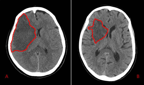
Methods: A cohort of ten patients with a first acute (within six hours since symptoms onset) ischemic stroke and a group of eight age- and sex-matched normal subjects were included. Groups were also matched for metabolic profile. All subjects underwent a blood sample collection for anandamide (AEA), 2-arachidonoylglycerol (2-AG) and palmitoylethanolamide (PEA) measurement; blood sampling was repeated in patients on admission (T0), at 6 (T1) and 18 hours (T2) thereafter. Patients neurological impairment was assessed using NIHSS and Fugl-Meyer Scale arm subitem (FMSa); stroke volume was determined on 48 h follow-up brain CT scans. Blood samples were analyzed by liquid chromatography-atmospheric pressure chemical ionization-mass spectrometry.
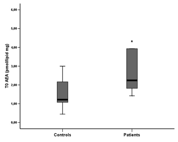
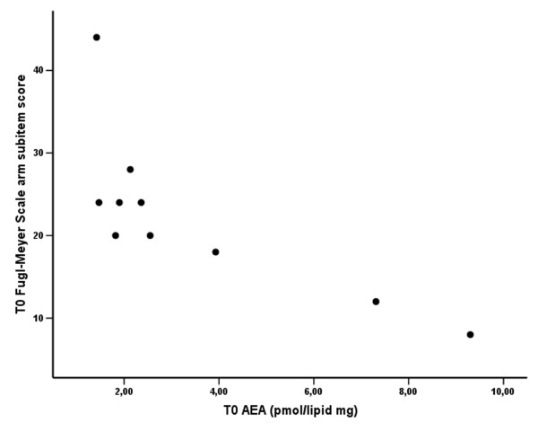
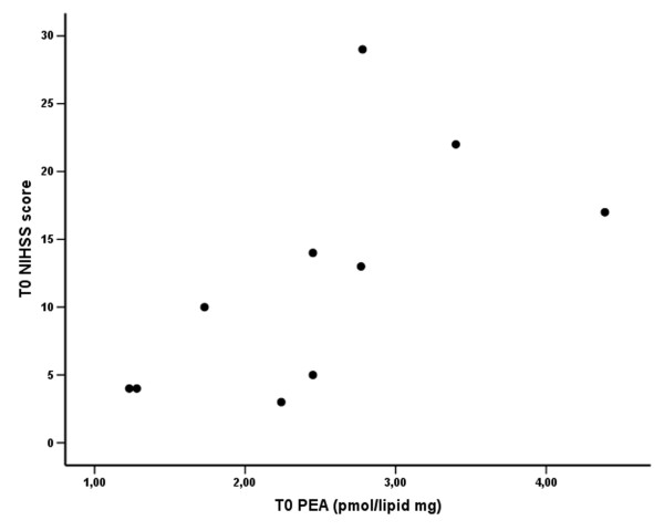
Results: 1)T0 AEA levels were significantly higher in stroke patients compared to controls. 2)A significant inverse correlation between T0 AEA levels and FMSa score was found. Moreover a positive correlation between T0 AEA levels and stroke volume were found in stroke patients. T0 PEA levels in stroke patients were not significantly different from the control group, but showed a significant correlation with the NIHSS scores. T0 2-AG levels were lower in stroke patients compared to controls, but such difference did not reach the significance threshold.
Conclusions: This is the first demonstration of elevated peripheral AEA levels in acute stroke patients. In agreement with previous murine studies, we found a significant relationship between AEA or PEA levels and neurological involvement, such that the greater the neurological impairment, the higher were these levels.
Figures
References
-
- Skaper SD, Buriani A, Dal Toso R, Petrelli L, Romanello S, Facci L, Leon A. The ALIamide palmitoylethanolamide and cannabinoids, but not anandamide, are protective in a delayed postglutamate paradigm of excitotoxic death in cerebellar granule neurons. Proc Natl Acad Sci USA. 1996;93:3984–3989. doi: 10.1073/pnas.93.9.3984. - DOI - PMC - PubMed
Publication types
MeSH terms
Substances
LinkOut - more resources
Full Text Sources
Other Literature Sources
Medical
