Review
. 2010 Jun;20(3):161-8.
doi: 10.1016/j.semcancer.2010.05.002. Epub 2010 May 12.

Matrix metalloproteinases (MMPs) and tissue inhibitors of metalloproteinases (TIMPs): Positive and negative regulators in tumor cell adhesion

Affiliations
Review

Matrix metalloproteinases (MMPs) and tissue inhibitors of metalloproteinases (TIMPs): Positive and negative regulators in tumor cell adhesion

Dimitra Bourboulia et al. Semin Cancer Biol. 2010 Jun.

Abstract

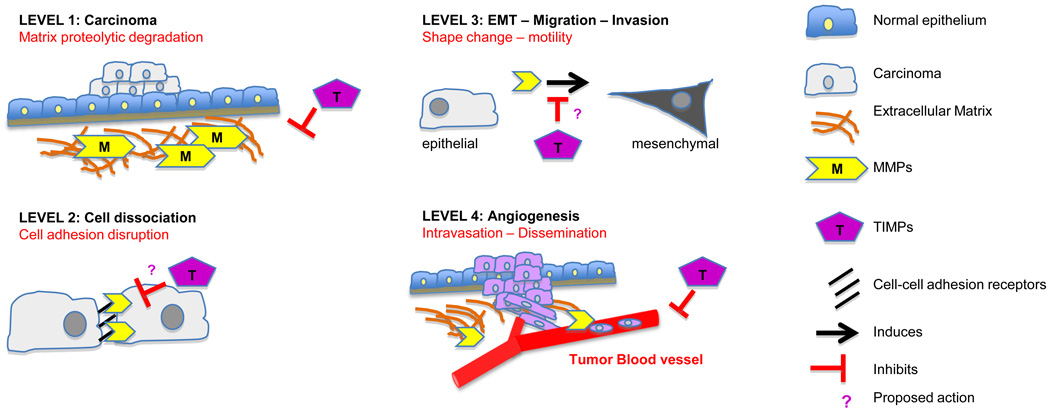
Cells adhere to one another and/or to matrices that surround them. Regulation of cell-cell (intercellular) and cell-matrix adhesion is tightly controlled in normal cells, however, defects in cell adhesion are common in the majority of human cancers. Multilateral communication among tumor cells with the extracellular matrix (ECM) and neighbor cells is accomplished through adhesion molecules, ECM components, proteolytic enzymes and their endogenous inhibitors. There is sufficient evidence to suggest that reduced adherence is a tumor cell property engaged during tumor progression. Tumor cells acquire the ability to change shape, detach and easily move through spaces disorganizing the normal tissue architecture. This property is due to changes in expression levels of adhesion molecules and/or due to elevated levels of secreted proteolytic enzymes, including matrix metalloproteinases (MMPs). Among other roles, MMPs degrade the ECM and, therefore, prepare the path for tumor cells to migrate, invade and spread to distant secondary areas, where they form metastasis. Tissue inhibitors of metalloproteinases or TIMPs control MMP activities and, therefore, minimize matrix degradation. Both MMPs and TIMPs are involved in tissue remodeling and decisively regulate tumor cell progression including tumor angiogenesis. In this review, we describe and discuss data that support the important role of MMPs and TIMPs in cancer cell adhesion and tumor progression.


Conflict of interest statement

Conflict of Interest

The authors declare that there is no conflict of interest.

Figures

Figure 1
Figure 1. Matrix Metaloproteinases (MMPs) domain structures and functions
There are 5 main MMP groups based on their structure and substrate: collagenases, gelatinases, matrilysins, membrane type MMPs (MT-MMPs) and stromelysins. The basic domain structure consists of: i) a signal peptide (SP) that directs the protein through the endoplasmic reticulum prior to secretion, ii) a pro-peptide region (PRO) that secures enzymatic latency and inactivity by blocking the catalytic site from becoming accessible to substrates or to inhibitors, iii) a catalytic region that, after the pro-peptide removal, confers the MMPs with their enzymatic activity with the help of zinc (Zn) ions and iv) a hemopexin domain at the C-terminus, that provides the specificity to and interaction with the substrates or the inhibitors (TIMPs), also presents the substrate to the catalytic domain via a high flexible hinge, except for matrilysins that lack the hemopexin-like and the hinge that joints it to the catalytic domain. The prodomain contains a cysteine-to-zinc switch motif where the Zn+2 at the catalytic domain binds to the cysteine at the prodomain and inhibits MMP activation and interaction with the substrate. The zinc in the catalytic domain interacts with the three histidines (H) in a motif sequence (HEXXHXXGXXH). Upon proteolytic processing and removal of the prodomain, the mature MMPs become active and able to catalyze. Although the majority of the MMPs are secreted as inactive zymogens, the MT-MMPs, MMP-11 and MMP-28 (epilysin) contain the furin-like motif RXKR between the proenzyme and catalytic domains allowing them to be activated by intracellular serine proteinases before they get secreted or localize to the cell membrane. The type I transmembrane (TM type I) MMPs are anchored to the membrane with a hydrophobic domain and a short cytoplasmic tail. The two exceptions are the MMP-17 and MMP-25 that instead attach with a glycosyl-phosphatidyl inositol (GPI) anchor domain. Gelatinases A and B or MMP-2 and MMP-9 also contain three collagen-binding fibronectin type II repeats within the catalytic domain. In addition, MMP-9 contains a serine-threonine and prolice rich O-glycosylated domain (O-glyco).
Figure 2
Figure 2. MMPs and TIMPs input in tumor cell adhesion
A schematic representation of MMPs and TIMPs role in cell adhesion modulation during different the levels of carcinoma progression (see text). Proposed (?) TIMPs actions remained to be discovered.

References

    1. Gumbiner BM. Cell adhesion: the molecular basis of tissue architecture and morphogenesis. Cell. 1996;84:345–357. - PubMed
    1. Nelson CM, Bissell MJ. Of extracellular matrix, scaffolds, and signaling: tissue architecture regulates development, homeostasis, and cancer. Annu Rev Cell Dev Biol. 2006;22:287–309. - PMC - PubMed
    1. Brooks SA, Lomax-Browne HJ, Carter TM, Kinch CE, Hall DM. Molecular interactions in cancer cell metastasis. Acta Histochem. 2010;112:3–25. - PubMed
    1. Christofori G. New signals from the invasive front. Nature. 2006;441:444–450. - PubMed
    1. Guan JL. Integrin signaling through FAK in the regulation of mammary stem cells and breast cancer. IUBMB Life. 2010 - PMC - PubMed

Publication types

MeSH terms

Substances