Review
. 2010 Jul-Aug;55(4):299-334.
doi: 10.1016/j.survophthal.2009.10.002. Epub 2010 May 14.

The neuro-ophthalmology of mitochondrial disease

Affiliations
Review

The neuro-ophthalmology of mitochondrial disease

J Alexander Fraser et al. Surv Ophthalmol. 2010 Jul-Aug.

Abstract

Mitochondrial diseases frequently manifest neuro-ophthalmologic symptoms and signs. Because of the predilection of mitochondrial disorders to involve the optic nerves, extraocular muscles, retina, and even the retrochiasmal visual pathways, the ophthalmologist is often the first physician to be consulted. Disorders caused by mitochondrial dysfunction can result from abnormalities in either the mitochondrial DNA or in nuclear genes which encode mitochondrial proteins. Inheritance of these mutations will follow patterns specific to their somatic or mitochondrial genetics. Genotype-phenotype correlations are inconstant, and considerable overlap may occur among these syndromes. The diagnostic approach to the patient with suspected mitochondrial disease entails a detailed personal and family history, careful ophthalmic, neurologic, and systemic examination, directed investigations, and attention to potentially life-threatening sequelae. Although curative treatments for mitochondrial disorders are currently lacking, exciting research advances are being made, particularly in the area of gene therapy. Leber hereditary optic neuropathy, with its window of opportunity for timely intervention and its accessibility to directed therapy, offers a unique model to study future therapeutic interventions. Most patients and their relatives benefit from informed genetic counseling.


Figures

Figure 1
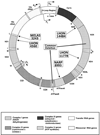
Figure 1. Map of the human mitochondrial genome
The human mitochondrial genome is comprised of 16,569 base pairs of nucleotides encoding 37 genes. Shown are the most frequent mtDNA point mutations responsible for mitochondrial disorders (Leber hereditary optic neuropathy (LHON); neurogenic muscle weakness, ataxia, and retinitis pigmentosa (NARP); and mitochondrial encephalomyopathy, lactic acidosis, and stroke-like episodes (MELAS)), and the 5 kilobase “common deletion” seen in chronic progressive external ophthalmoplegia (CPEO). (Adapted from: www.mitomap.org).
Figure 2
Figure 2. Nuclear and mitochondrial DNA influences on mitochondrial function
Normal mitochondrial function depends on the symbiotic relationship of nuclear (blue) and mitochondrial (red) DNA influences. Normal cellular homeostatic processes (arrows with plain text) are under the dual control of these two genomes (red arrows: mitochondrial-DNA-related processes; black arrows: nuclear-DNA-mediated extramitochondrial processes; blue arrows: nuclear-DNA-mediated intramitochondrial processes). Mitochondrial diseases (underlined text) may therefore arise from dysfunctional homeostatic processes mediated by either genome (red underlined text: diseases of mitochondrial DNA origin; blue underlined text: diseases of nuclear DNA origin). The mitochondrial genome (dark red circle) encodes 13 structural subunits of the OXPHOS machinery (complexes I, III, IV, and V) (light red semicircles) and 24 molecules required for mitochondrial gene translation (2 rRNAs (light red ovals) and 12 tRNAs (light red cloverleaf)). The nuclear genome (paired dark blue lines) encodes all other proteins used by mitochondria. Nuclear proteins (light blue cloverleaf) imported into the mitochondrial matrix comprise the remaining structural subunits of the OXPHOS machinery (dark blue semicircles and circles) and are involved in: the assembly and repair of the OXPHOS complexes; the fusion of mitochondria within the cytosol; the detoxification of reactive oxygen species; the stabilization of mitochondrial membranes; the sequestration of the pro-apoptotic molecule cytochrome c; and the repair, replication, and expression of mitochondrial genes (blue arrows pointing to red arrows). Abbreviations: OXPHOS: oxidative phosphorylation system (comprised of complexes I to V) ATP: adenosine triphosphate ROS: reactive oxygen species nP: nuclear product (proteins) mP: mitochondrial product (proteins, tRNAs, and rRNAs) nDNA: nuclear DNA mtDNA: mitochondrial DNA mRNA: messenger RNA tRNA: transfer RNA rRNA: ribosomal RNA DOA: dominant optic atrophy OPA1+: “OPA1-Plus” syndrome adCPEO: autosomal dominant chronic progressive external ophthalmoplegia MNGIE: mitochondrial neurogastrointestinal encephalomyopathy CMT2A: Charcot-Marie-Tooth disease, type 2A FRDA: Friedreich ataxia sCPEO: sporadic chronic progressive external ophthalmoplegia miCPEO: maternally inherited chronic progressive external ophthalmoplegia LHON: Leber hereditary optic neuropathy MELAS: mitochondrial encephalomyopathy, lactic acidosis, and stroke-like episodes NARP: neurogenic muscle weakness, ataxia, and retinitis pigmentosa
Figure 3
Figure 3. Modes of genetic inheritance
(A) Maternal inheritance (e.g., Leber hereditary optic neuropathy (LHON)): women transmit the mutation to all children, while men do not transmit the mutation to any children; (B) autosomal dominant inheritance (e.g., dominant optic atrophy (DOA)): men and women alike have a 50% chance of transmitting the mutation to each child; (C) autosomal recessive inheritance (e.g., Friedreich ataxia (FRDA)): each parent’s mutant allele has a 50% chance of being transmitted to the next generation, but disease is only expressed when a child receives an allele from both parents.
Figure 4
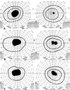
Figure 4. Goldmann visual fields (GVF) in a patient with Leber hereditary optic neuropathy (LHON)
Progressive central scotoma in the right eye of a 21 year old man with the 11778 LHON mutation. He had suffered a painless central scotoma in the left eye three months prior. (A) GVF from March 5, 2009; (B) GVF from March 19. 2009; (C) GVF from April 16, 2009.
Figure 5
Figure 5. Ocular fundus appearance in a patient with Leber hereditary optic neuropathy (LHON)
(A) Acute pattern: disc hyperemia, pseudoedema, and telangiectasias; the left eye was affected one month prior to the right eye, and early temporal optic disc pallor is evident in the left eye. (B) Chronic pattern: diffuse optic atrophy, most apparent temporally, three years later.
Figure 6
Figure 6. Primary and secondary LHON mutations
Primary LHON point mutations (inside circular mitochondrial genome) and secondary LHON point mutations (outside circular mitochondrial genome) are shown. Mutations marked * may be primary, but they account for only one or a few pedigrees worldwide. Mutations marked *d are primary mutations associated with LHON and dystonia; mutations marked *m are primary mutations associated with LHON and MELAS; mutation marked *e is a primary mutation associated with LHON and encephalopathy. (Adapted from: www.mitomap.org).
Figure 7
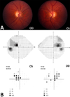
Figure 7. Ocular fundus appearance and visual fields in a patient with dominant optic atrophy (DOA)
(A) Classic “temporal wedge” of optic disc pallor, symmetric between eyes; (B) Humphrey visual fields showing bilateral cecocentral scotomas in grayscale (top) and pattern deviation (bottom), reflecting involvement of the papillomacular bundles OU. Note the superobitemporal predilection of the visual field defecst.
Figure 8
Figure 8. External motility photographs in chronic progressive external ophthalmoplegia
(A) Bilateral ptosis and bifacial weakness; (B) Limitation of ocular motility in all directions in both eyes.
Figure 9
Figure 9. Classic histopathological findings in mitochondrial myopathy
(A) Gomori trichrome stain: ragged red fibers (RRFs); (B) Cytochrome oxidase (COX) stain: isolated COX-negative fiber (center); (C) Succinate dehydrogenase (SDH) stain: increased staining of a blood vessel (arrow) in a patient with MELAS; (D) Electron microscopy: paracrystalline “parking lot” inclusions. (Images reproduced with permission from: Bourgeois JM, Tarnopolsky MA. Pathology of skeletal muscle in mitochondrial disorders. Mitochondrion. 2004;4(5–6):441-42).
Figure 10
Figure 10. Ocular fundus appearance in a patient with pigmentary retinopathy associated with Kearns-Sayre syndrome (KSS)
(A) Diffuse retinal arteriolar attenuation and mild waxy pallor of the optic disc; (B) salt and pepper retinopathy.
Figure 11
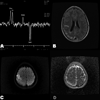
Figure 11. Neuroradiological features of MELAS
(A) Magnetic resonance spectroscopy (MRS) showing a decreased N-acetyl aspartate (NAA) peak relative to choline (Cho) and creatine (Cr) and a large lactate doublet (Lact); (B) Fluid-attenuated inversion recovery (FLAIR) imaging sequence showing high T2 signal in a cortical laminar distribution of the right brain, predominantly posteriorly, sparing deep white matter, and not respecting cerebral arterial territories; (C) Diffusion weighted imaging (DWI) sequence, showing a right cortical focus of restricted diffusion during a stroke-like episode; (D) Apparent diffusion coefficient (ADC) map in the same patient as (C), showing no decreased signal in the involved area of cortex. (Images courtesy of Dr. Chad Holder, Emory University).

References

    1. Abe K, Yoshimura H, Tanaka H, et al. Comparison of conventional and diffusion-weighted MRI and proton MR spectroscopy in patients with mitochondrial encephalopathy, lactic acidosis, and stroke-like events. Neuroradiology. 2004;46:113–117. - PubMed
    1. Adjadj E, Mansouri K, Borruat FX. Mitochondrial DNA (mtDNA) A3243G mutation associated with an annular perimacular retinal atrophy. Klin Monatsbl Augenheilkd. 2008;225:462–464. - PubMed
    1. Ahn J, Kim NJ, Choung HK, et al. Frontalis sling operation using silicone rod for the correction of ptosis in chronic progressive external ophthalmoplegia. Br J Ophthalmol. 2008;92:1685–1688. - PubMed
    1. Aijaz S, Erskine L, Jeffery G, et al. Developmental expression profile of the optic atrophy gene product: OPA1 is not localized exclusively in the mammalian retinal ganglion cell layer. Invest Ophthalmol Visual Sci. 2004;45:1667–1673. - PubMed
    1. Amati-Bonneau P, Milea D, Bonneau D, et al. OPA1-associated disorders: phenotypes and pathophysiology. Int J Biochem Cell Biol. 2009;41:1855–1865. - PubMed

Publication types

Substances