. 2010 May 28;32(5):616-27.
doi: 10.1016/j.immuni.2010.04.016. Epub 2010 May 13.

Epigenetic instability of cytokine and transcription factor gene loci underlies plasticity of the T helper 17 cell lineage

Affiliations

Epigenetic instability of cytokine and transcription factor gene loci underlies plasticity of the T helper 17 cell lineage

Ryuta Mukasa et al. Immunity. .

Abstract

Phenotypic plasticity of T helper 17 (Th17) cells suggests instability of chromatin structure of key genes of this lineage. We identified epigenetic modifications across the clustered Il17a and Il17f and the Ifng loci before and after differential IL-12 or TGF-beta cytokine signaling, which induce divergent fates of Th17 cell precursors. We found that Th17 cell precursors had substantial remodeling of the Ifng locus, but underwent critical additional modifications to enable high expression when stimulated by IL-12. Permissive modifications across the Il17a-Il17f locus were amplified by TGF-beta signaling in Th17 cells, but were rapidly reversed downstream of IL-12-induced silencing of the Rorc gene by the transcription factors STAT4 and T-bet. These findings reveal substantial chromatin instability of key transcription factor and cytokine genes of Th17 cells and support a model of Th17 cell lineage plasticity in which cell-extrinsic factors modulate Th17 cell fates through differential effects on the epigenetic status of Th17 cell lineage factors.


Figures

Figure 1
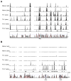
Figure 1. DNase I HS maps of the Ifng and Il17a-Il17f loci
Naïve CD4+ T cells from OT-II TCR transgenic mice were isolated and cultured under Th1- or Th17-polarizing conditions. Cells were harvested without (rest) or with (stim) PMA+ionomycin stimulation and subjected to DNase I digestion and DNase-chip analysis. Panels show results of Ifng (A) and Il17a-Il17f (B) loci, respectively, aligned with corresponding VISTA plot (Frazer et al., 2004), where mouse sequence is shown on the x axis and percentage similarity to human on the y axis.
Figure 2
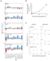
Figure 2. IL-12 and TGF β induce differential cytokine expression phenotype in Th17 precursor cells
(A) CD4+ T cells from OT-II mice were cultured under Th1 or Th17 cell-polarizing conditions. Th17 cells were harvested and restimulated in the presence of TGFβ or IL-12, anti-IL-4 and OVAp for an additional 6 days. Cells were stained intracellularly for IL-17A and IFNγ after PMA+ionomycin activation. Data in the quadrants are the frequencies of CD4+ cells. (B) Cumulative data for frequencies of cytokine-positive CD4+ T cells analyzed as (A). Th1 and Th17 cells were harvested on day5 for ChIP analysis. For Th17+IL-12 and Th17+TGFβ cells, Th17 cells were harvested on day 6 or 7 of the first polarization and restimulated in the presence of IL-12 or TGFβ for another 6 days (means ± SD).
Figure 3
Figure 3. Th17 cells undergo rapid epigenetic remodeling and acquire Th1-like histone modifications across the Ifng locus following restimulation with IL-12
(A) Th1, Th17, Th17+IL-12 and Th17+TGF β cells were derived from CD4+ T cells of OT-II TCR transgenic mice as in Figure 2. Cells were then processed for ChIP analysis using antibodies specific for H3K4me or H3K27me3. Data for H3K4me and H3K27me3 were normalized against the value for naïve CD4+ T cells and input DNA, respectively (means ± SD). (B) CD4+ T cells from OT-II TCR transgenic mice were cultured under Th17-polarizing conditions. Recovered cells were restimulated in the presence of anti-IL-4 and TGF® or IL-12 for the indicated times and processed for expression of Ifng mRNA after 2 h of PMA+ionomycin activation (means ± SD). (C) Th17 cells restimulated with anti-CD3 and anti-CD28 in the presence of IL-12 and anti-IL-4 for 6, 24, or 48 hr as in (B). Cells were processed for ChIP analysis with antibodies specific for H3K4me or H3K27me3. Data are normalized against cells without restimulation (0 hr; means ± SD).
Figure 4
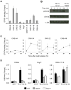
Figure 4. STAT4 and T-bet are necessary for epigenetic remodeling of the Ifng locus following IL-12 stimulation of Th17 cells
(A) OT-II Th17 cells were restimulated with anti-CD3 and anti-CD28 in the presence of IL-12 and anti-IL-4 for 24 hr and processed for STAT4 ChIP analysis. Data are normalized against input DNA (means ± SD). (B) Th17 cells were derived from Il17fThy1.1/Thy1.1 mice and Thy1.1+ (IL-17F+) Th17 cells isolated by magnetic sorting. Cells were restimulated with anti-CD3 and anti-CD28 in the presence of IL-12 or TGF® for the indicated times, and immunoblot analysis was performed on isolated nuclei using antibodies specific for phopho-STAT4, total STAT4 or control (c-Jun). (C) Th17 cells were restimulated with anti-CD3 and anti-CD28 in the presence of IL-12 and anti-IL-4 for 6, 24, or 48 hr and cells were processed for STAT4 ChIP analysis as in (A). Data are normalized against input DNA and represent the mean ± SD of pooled data from two to four separate experiments. (D) Th17 cells were derived from wild type (WT), Stat4-/-, and Tbx21-/- mice and restimulated with IL-12, anti-CD3, anti-IL-4, and anti-IFN© for 6 days. Th17 presursor cells (Th17) and cells recovered after 6 days restimulation with IL-12 (Th17+IL-12) were processed for H3K4me ChIP. Data are normalized against input DNA (means ± SD).
Figure 5
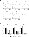
Figure 5. IL-12 induces repressive histone modification at Il17a-Il17f locus of Th17 cells
(A) Naïve, Th1, Th17, Th17+IL-12, Th17+TGF® cells were derived from OT-II CD4+ T cells as in Figure 3A then analyzed for H3K4me and H3K27me3 modification of the indicated sites by ChIP. Data are normalized against input DNA (means ± SD). (B) OT-II Th17 cells were restimulated with anti-CD3 and anti-CD28 in the presence of anti-IL-4 and TGF® or IL-12 for the indicated times and processed for expression of Il17a mRNA after 2 hr of activation with PMA+ionomycin (means ± SD). (C) Th17 cells were restimulated with anti-CD3 and anti-CD28 in the presence of IL-12 and anti-IL-4 for 6, 24, or 48 h as in (B). Cells were then processed for ChIP assay with antibody specific for H3K4me or H3K27me3. Data are normalized against cells without restimulation (0 hr) and represent means ± SD.
Figure 6
Figure 6. IL-12-induced extinction of the Il17a-Il17f locus in Th17 cells is associated with rapid modulation of expression of Th17- and Th1-lineage transcription factors
(A) Pure Th17 cells (Thy1.1+) were derived CD4+ T cells of from Il17fThy1.1/Thy1.1 mice and were restimulated with IL-12 or TGF® for 1, 3, or 5 days and processed for quantification of mRNA of the indicated genes by RT-PCR. Data are normalized to 18S rRNA and expressed as relative values to Thy1.1+ cells used before restimulation. The horizontal dotted lines indicate the values of each mRNA in naïve CD4+ T cells from Il17fThy1.1/Thy1.1 mice. Data are representative of two independent experiments. (B) Th17 precursor cells prepared from wild type (WT), Stat4-/-, and Tbx21-/- mice were harvested and divided into two fractions; one (Th17) processed for without further treatment for mRNA quantitation of the indicated transcripts, and the other (Th17+IL-12) restimulated with IL-12, anti-IL-4, and anti-IFN© for 6 days and processed identically. Data are normalized to ®2-microglobulin and expressed as relative values to WT Th17 cells (mean ± SD) and are representative of two similar experiments.
Figure 7
Figure 7. Extinction of the Il17a-Il17f locus is linked to STAT4- and T-bet-induced epigenetic repression of the Rorc gene
(A) CD4+ T cells isolated from Il17fThy1.1/Thy1.1 mice were activated with anti-CD3+CD28 coated beads under Th17 cell-polarizing conditions on day 0 and were transduced with the retroviral vectors encoding IRES-GFP (GFP (control)) or ROR©t-IRES-GFP (ROR©t-GFP) on day 1. Th17 cells were harvested on day 6 and a fraction of cells were stained for Thy1.1 and intracellular IL-17A and IFN© after PMA+ionomycin activation. Thy1.1+ (IL-17F+) cells were isolated from a second fraction by magnetic sorting and restimulated with anti-CD3 and anti-CD28 in the presence of anti-IL-4, anti-IFN©, and IL-12 for 6 days. Recovered Th17+IL-12 cells were analyzed for intracellular IL-17A and IFN© after PMA+ionomycin activation. Thy1.1+GFP+ cells and GFP+ cells were gated for analysis of Th17 and Th17+IL-12 cells, respectively. (B) CD4+ T cells of Il17fThy1.1/Thy1.1 mice were cultured under Th17 cell-polarizing conditions in the presence of anti-IL-12p40 for 6 days, and Thy1.1+ (IL-17F+) cells were isolated by magnetic sorting. ChIP for H3K4me and H3K27me3 histone modifications was performed on this fraction of the isolated Thy1.1+ cells (Th17). Th17+IL-12 and Th17+TGFβ cells were generated from a second fraction of Thy1.1+ cells and also evaluated for H3K4me and H3K27me3 by ChIP. Data are normalized against input DNA (mean ± SD). (C) Th17 and Th17+IL-12 cells were derived from wild type (WT), Stat4-/-, and Tbx21-/- mice as in (B) and analyzed for H3K27me3 modification of Rorc+5.0 by ChIP. Data are normalized against input DNA (mean ± SD). (D) Th17+IL-12 and Th17+TGFβ cells were generated from Il17fThy1.1/Thy1.1 CD4+ T cells as in (B) and analyzed for binding of T-bet to the indicated sites in the Rorc locus by T-bet-specific ChIP. Data are normalized against input DNA (mean ± SD).

Comment in

  • Keeping one's option open.
    Collins PL, Aune TM. Collins PL, et al. Immunity. 2010 May 28;32(5):581-3. doi: 10.1016/j.immuni.2010.05.008. Immunity. 2010. PMID: 20510865

References

    1. Akimzhanov AM, Yang XO, Dong C. Chromatin remodeling of interleukin-17 (IL-17)-IL-17F cytokine gene locus during inflammatory helper T cell differentiation. J Biol Chem. 2007;282:5969–5972. - PubMed
    1. Ansel KM, Djuretic I, Tanasa B, Rao A. Regulation of Th2 differentiation and Il4 locus accessibility. Annu Rev Immunol. 2006;24:607–656. - PubMed
    1. Ansel KM, Lee DU, Rao A. An epigenetic view of helper T cell differentiation. Nat Immunol. 2003;4:616–623. - PubMed
    1. Bending D, De La Pena H, Veldhoen M, Phillips JM, Uyttenhove C, Stockinger B, Cooke A. Highly purified Th17 cells from BDC2.5NOD mice convert into Th1-like cells in NOD/SCID recipient mice. J Clin Invest. 2009;119:565–572. - PMC - PubMed
    1. Chang S, Aune TM. Histone hyperacetylated domains across the Ifng gene region in natural killer cells and T cells. Proc Natl Acad Sci U S A. 2005;102:17095–17100. - PMC - PubMed

Publication types

MeSH terms