Review
. 2010 May 13;66(3):337-51.
doi: 10.1016/j.neuron.2010.04.028.

Unraveling mechanisms of homeostatic synaptic plasticity

Affiliations
Review

Unraveling mechanisms of homeostatic synaptic plasticity

Karine Pozo et al. Neuron. .

Abstract

Homeostatic synaptic plasticity is a negative feedback mechanism that neurons use to offset excessive excitation or inhibition by adjusting their synaptic strengths. Recent findings reveal a complex web of signaling processes involved in this compensatory form of synaptic strength regulation, and in contrast to the popular view of homeostatic plasticity as a slow, global phenomenon, neurons may also rapidly tune the efficacy of individual synapses on demand. Here we review our current understanding of cellular and molecular mechanisms of homeostatic synaptic plasticity.


Figures

Figure 1
Figure 1. Basic scheme of homeostatic synaptic plasticity at an excitatory synapse
a. Basal conditions: Synaptic transmission is mediated via the liberation of neurotransmitters from the presynaptic terminal and subsequent activation of receptors on the postsynaptic cell. The efficacies of neurotransmitter release and reception are major determinants of pre- and postsynaptic strengths, respectively. b,c. Neurons offset imposed changes in network activity by adapting their pre and postsynaptic strengths. (b) Reduced activity is offset presynaptically by enhancing the recycling of vesicles, the number of docked vesicles, and the release probability. Postsynaptically, additional neurotransmitter receptors are incorporated at the synapse by a mechanism involving lateral diffusion from extrasynaptic sites and exocytosis from intracellular pools. (c) To compensate an increased network activity, presynaptic neurons decrease their release probability while postsynaptic cells reduce the number of postsynaptic receptors by endocytosis or by lateral diffusion from synaptic to extrasynaptic sites. Depending on the developmental stage and on experimental conditions, pre- and postsynaptic changes can occur concurrently or separately. Glial cells (shown in grey) can also contribute to the changes in synaptic strength, for example, by secreting soluble factors that signal through cell surface receptors. See text for details.
Figure 2
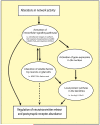
Figure 2. Summary of the molecular mechanisms underlying homeostatic synaptic plasticity
Changes in network activity are detected by an unknown mechanism by neurons or glial cells and activate intracellular mechanisms to modify presynaptic release and/or the abundance of functional postsynaptic receptors. This can involve activation of gene expression in neurons and triggering of local dendritic protein synthesis (e.g. Arc/Arg3.1, AMPA receptors) as well as the release of soluble factors such as BDNF or TNFα from neurons and glial cells that then engage additional signaling pathways. See text for details.

References

    1. Aakalu G, Smith WB, Nguyen N, Jiang C, Schuman EM. Dynamic Visualization of Local Protein Synthesis in Hippocampal Neurons. Neuron. 2001;30:489–502. - PubMed
    1. Abraham W, Goddard G. Asymmetric relationships between homosynaptic long-term potentiation and heterosynaptic long-term depression. Nature. 1983;305:717–719. - PubMed
    1. Aoto J, Nam CI, Poon MM, Ting P, Chen L. Synaptic Signaling by All-Trans Retinoic Acid in Homeostatic Synaptic Plasticity. Neuron. 2008;60:308–320. - PMC - PubMed
    1. Aptowicz CO, Kunkler PE, Kraig RP. Homeostatic plasticity in hippocampal slice cultures involves changes in voltage-gated Na+ channel expression. Brain Res. 2004;998:155–163. - PMC - PubMed
    1. Armstrong JN, Saganich MJ, Xu N-J, Henkemeyer M, Heinemann SF, Contractor A. B-Ephrin Reverse Signaling Is Required for NMDA-Independent Long-Term Potentiation of Mossy Fibers in the Hippocampus. J Neurosci. 2006;26:3474–3481. - PMC - PubMed

Publication types

LinkOut - more resources