. 2010 May 13;66(3):417-28.
doi: 10.1016/j.neuron.2010.04.037.

Distinct contributions of rod, cone, and melanopsin photoreceptors to encoding irradiance

Affiliations

Distinct contributions of rod, cone, and melanopsin photoreceptors to encoding irradiance

Gurprit S Lall et al. Neuron. .

Abstract

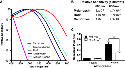
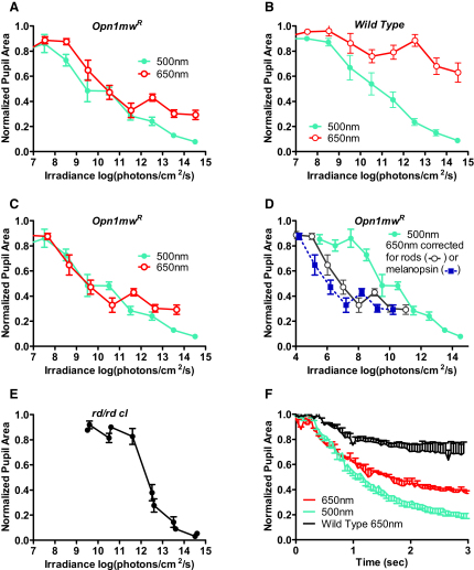
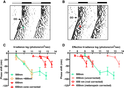
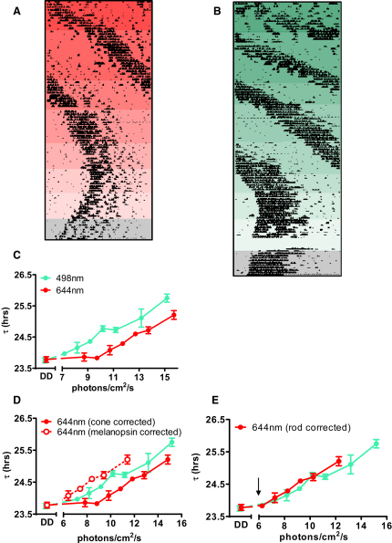
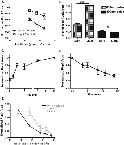
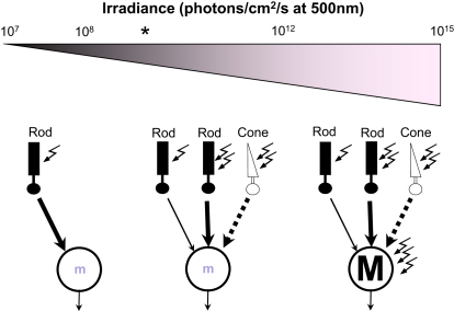
Photoreceptive, melanopsin-expressing retinal ganglion cells (mRGCs) encode ambient light (irradiance) for the circadian clock, the pupillomotor system, and other influential behavioral/physiological responses. mRGCs are activated both by their intrinsic phototransduction cascade and by the rods and cones. However, the individual contribution of each photoreceptor class to irradiance responses remains unclear. We address this deficit using mice expressing human red cone opsin, in which rod-, cone-, and melanopsin-dependent responses can be identified by their distinct spectral sensitivity. Our data reveal an unexpectedly important role for rods. These photoreceptors define circadian responses at very dim "scotopic" light levels but also at irradiances at which pattern vision relies heavily on cones. By contrast, cone input to irradiance responses dissipates following light adaptation to the extent that these receptors make a very limited contribution to circadian and pupillary light responses under these conditions. Our data provide new insight into retinal circuitry upstream of mRGCs and optimal stimuli for eliciting irradiance responses.


Figures

Figure 1
Figure 1
Enhanced Long-Wavelength Sensitivity of Cone Vision in Opn1mwR Mice (A) In Opn1mwR mice, the native mouse m-cone opsin (dotted green line, shows spectral sensitivity approximated by opsin nomogram [Govardovskii et al., 2000] with peak sensitivity [λmax] = 511 nm) is lost and replaced with a human red cone opsin (Smallwood et al., 2003) whose spectral sensitivity (red line; λmax = 556 nm) profile is quite distinct from that of mouse rod (λmax = 498 nm), melanopsin (λmax = 480 nm), and s-cone (λmax = 360 nm) opsins (black, blue, and purple lines, respectively). (B) Divergence in the spectral sensitivity of red cones, rods, and melanopsin is reflected in large differences in their relative sensitivity to mid- (500 nm) and long- (600 or 650 nm) wavelength light. (C) Red cone input to the pupil light reflex is revealed as a significant increase in response to a 1 min, 650 nm stimulus (3 × 10¹⁴ photons/cm2/s) in Opn1mwR mice compared with wild-type mice (mean ± SEM; t test, p < 0.01). The genotypes showed similar responses to an equivalent (1 min; 3 × 10¹⁴ photons/cm2/s) 500 nm stimulus (mean ± SEM; n = 8–12; t test, p > 0.05).
Figure 2
Figure 2
Cone Contributions to Defining Pupil Size (A) Sixty-second stimuli drove irradiance-dependent decreases in pupil size at both 500 and 650 nm in dark-adapted Opn1mwR mice (n = 4–12). (B) Wild-type mice also responded to both wavelengths, but were much less sensitive to 650 than 500 nm (n = 4–5). (C) Following correction of the 650 nm data from Opn1mwR mice to allow for the reduced sensitivity of cones to this wavelength versus 500 nm (irradiance of 650 nm stimuli × 0.13), the two irradiance response curves are superimposed at <1011 photons/cm2/s. F-test analysis reveals that sigmoidal curves fitted to these data sets differ (p < 0.01) in the best fit for their lower asymptote (nonoverlapping 95% confidence intervals), but not in other parameters. (D) There was no such convergence when irradiances were normalized according to the spectral sensitivity of rods or melanopsin. (E) As previously reported (Lucas et al., 2003), the pupillary responses of dark-adapted rd/rd cl mice elicited by 60 s 500 nm stimuli had a high threshold (n = 4–5). (F) A detailed examination of pupil size (n = 4) over the first 3 s of exposure (lights on at time = 0) to bright stimuli at 500 nm (2.2 × 10¹³ photons/cm2/s) and 650 nm (1.6 × 10¹⁴ photons/cm2/s) isoluminant for red cones revealed that, whereas the early rate of constriction was equivalent, responses at the two wavelengths diverged after ∼0.8 s. The response of wild-type mice to the longer wavelength was smaller and slower, confirming that the 650 nm response over these timescales relies on red cones. All data points show mean ± SEM pupil area normalized to prestimulus condition.
Figure 3
Figure 3
Rods Define Circadian Responses at Moderate Irradiances Representative double plotted actograms of wheel-running activity from the same Opn1mwR mouse exposed to 15 min pulses of 500 nm (1011 photons/cm2/s; A) or 650 nm (1012 photons/cm2/s; B) light at CT16 show a marked phase delay to the shorter, but not the longer, wavelength. The first 10–12 days show stable entrainment to a 12 hr:12 hr LD cycle (depicted as open/closed bars at the top of each panel), before release into DD at time indicated by arrow. Green/red circles represent the time of light exposure, with lines drawn through activity onsets before and after this pulse revealing the magnitude of the phase shift. (C) Irradiance response curves for phase delays elicited (mean ± SEM; n = 3–8) by 15 min pulses at CT16 using this paradigm in Opn1mwR mice reveal substantially reduced sensitivity to 650 nm light compared with 500 nm. Correcting for the relative sensitivity of cones at this wavelength was insufficient to account for the poor long-wavelength sensitivity. Responses to 500 nm were not altered by inclusion of 600 nm light at 10× higher irradiance (500+600 nm curve), confirming that cones make neither a strong stimulatory nor inhibitory contribution to this response. (D) Correcting the 650 nm irradiance response curve for the relative sensitivity of rods or melanopsin at this wavelength suggests that rods define the sensitivity of this response.
Figure 4
Figure 4
Responses to Constant Light Reveal a High Sensitivity Rod Input to the Circadian Clock (A and B) Representative actograms of wheel-running activity reveal irradiance-dependent increases in circadian period (τ) of Opn1mwR mice exposed to constant 644 nm (A) or 498 nm (B) light. Consecutive reductions in light intensity are depicted as reductions in background colored shading. The final 10 days of both records (gray shading) were collected in DD. (C) Irradiance response relationships for τ (estimated by periodogram analysis of activity records as shown in A and B) at 498 and 644 nm (mean ± SEM; n = 3–6), revealed substantially reduced sensitivity at the longer wavelength. (D) The difference in responsiveness to these two wavelengths could not be adequately accounted for by correcting for the relative sensitivity of either melanopsin or cones. (E) On the other hand, the curves became superimposed when corrected for rods. Arrow represents the estimated threshold for rod vision in mice (Nathan et al., 2006).
Figure 5
Figure 5
Cone Input to the PLR Is Reduced under Light-Adapted Conditions (A) Pre-exposure to 5 min 644 nm (1013 photons/cm2/s) substantially reduced pupillary responses (mean ± SEM; n = 6–7) to 10 s stimuli of equivalent or even higher irradiance (open circles) compared to those obtained following 1 hr of dark adaptation (closed circles). (B) Under more extensive adaptation (15 min 1.2 × 1015 photons/cm2/s; “Light”), pupil responses were completely absent throughout 30 s of exposure to a 650 nm test stimulus (2 × 1014 photons/cm2/s; hatched columns) capable of driving strong constriction under dark-adapted conditions (“Dark”). By contrast the response to an equivalent 500 nm (2 × 10¹³ photons/cm2/s; solid columns) test stimulus was unaffected. One-way ANOVA, p < 0.0001; selected post hoc tests with Tukey's correction shown, ∗∗∗p < 0.001; ns, p > 0.05. (C) The degree to which responses to the 30 s 650 nm test stimulus were inhibited was dependent upon the duration of prior light exposure (one-way ANOVA, p < 0.0001), although all exposures >15 s significantly impaired responses when compared with those of dark-adapted animals (Dunnett's post hoc comparisons, p < 0.01; n = 5–7). (D) Responsiveness to a 15 s 650 nm test stimulus (1.8 × 10¹⁴ photons/cm2/s) recovered over the course of 1 hr of dark adaptation (one-way ANOVA, p < 0.001; Dunnett's post hoc comparisons, p < 0.05, versus 0.1 min dark adaptation for times >1 min; n = 5–7). (E) Pre-exposure of rd/rd cl mice to 500 nm light (1.4 × 10¹⁴ photons/cm2/s) for either 5 or 60 min induced more classical light adaptation comprising a simple reduction in sensitivity to a subsequent 10 s 500 nm test pulse. All data points show mean ± SEM pupil area normalized to prestimulus condition.
Figure 6
Figure 6
Temporal Contrast Reveals Cone Input to the Circadian Clock (A) Phase delays (mean ± SEM; n = 3–8) in Opn1mwR mice exposed to 500 nm at CT16 presented as 15 × 1 min pulses over 43 min are indistinguishable from those elicited by continuous 15 min exposure at this wavelength. (B) In contrast, at 650 nm the discontinuous stimuli were substantially more efficient than equiquantal continuous pulses at eliciting phase shifts (mean ± SEM; n = 5–9).
Figure 7
Figure 7
These Data Suggest an Irradiance Measurement System with Four Distinct Inputs At very low irradiances responses rely solely on rods, probably signaling via the highest-sensitivity rod visual pathways. Over a broad range of moderate irradiances, lower-sensitivity rod and cone pathways take over. Under dark-adapted conditions cones dominate NIF responses, but their influence is reduced under light adaptation. This suggests that under most field conditions cones are restricted to contributing high-frequency modulation of pupil size with rather little influence on systems such as the circadian clock that integrate light signals over prolonged timescales. Under these circumstances, currently undefined rod pathways play the major role. Melanopsin phototransduction has low sensitivity, allowing it to encode high irradiances. Our data suggest thresholds of ∼107, 108, and 1012 500 nm photons/cm2/s for rod, cone, and melanopsin inputs, respectively. Asterisk depicts approximate threshold for murine cone-based vision for reference (Nathan et al., 2006).

References

    1. Abd-El-Barr M.M., Pennesi M.E., Saszik S.M., Barrow A.J., Lem J., Bramblett D.E., Paul D.L., Frishman L.J., Wu S.M. Genetic Dissection of Rod and Cone Pathways in the Dark-Adapted Mouse Retina. J. Neurophysiol. 2009;102:1945–1955. - PMC - PubMed
    1. Aggelopoulos N.C., Meissl H. Responses of neurones of the rat suprachiasmatic nucleus to retinal illumination under photopic and scotopic conditions. J. Physiol. 2000;523:211–222. - PMC - PubMed
    1. Alpern M. Rhodopsin kinetics in the human eye. J. Physiol. 1971;217:447–471. - PMC - PubMed
    1. Altimus C.M., Güler A.D., Villa K.L., McNeill D.S., Legates T.A., Hattar S. Rods-cones and melanopsin detect light and dark to modulate sleep independent of image formation. Proc. Natl. Acad. Sci. USA. 2008;105:19998–20003. - PMC - PubMed
    1. Bachleitner W., Kempinger L., Wülbeck C., Rieger D., Helfrich-Förster C. Moonlight shifts the endogenous clock of Drosophila melanogaster. Proc. Natl. Acad. Sci. USA. 2007;104:3538–3543. - PMC - PubMed

Publication types

LinkOut - more resources