. 2010 May 13;66(3):461-9.
doi: 10.1016/j.neuron.2010.03.034.

Normal movement selectivity in autism

Affiliations

Normal movement selectivity in autism

Ilan Dinstein et al. Neuron. .

Abstract

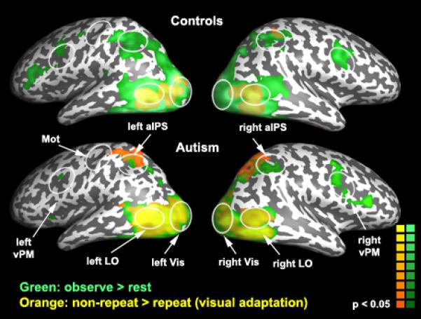
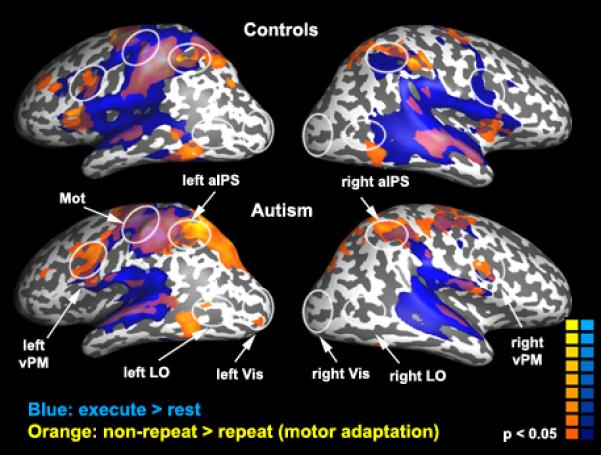
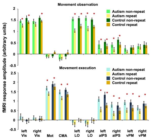
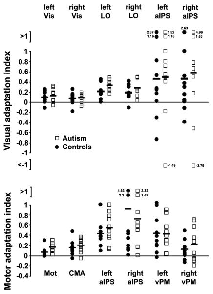
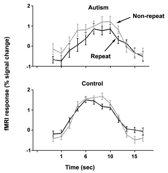
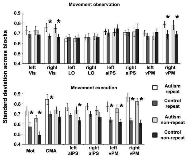
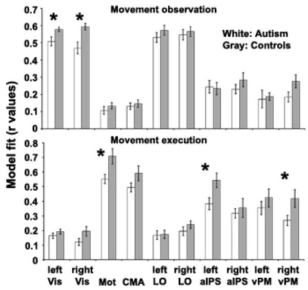
It has been proposed that individuals with autism have difficulties understanding the goals and intentions of others because of a fundamental dysfunction in the mirror neuron system. Here, however, we show that individuals with autism exhibited not only normal fMRI responses in mirror system areas during observation and execution of hand movements but also exhibited typical movement-selective adaptation (repetition suppression) when observing or executing the same movement repeatedly. Movement selectivity is a defining characteristic of neurons involved in movement perception, including mirror neurons, and, as such, these findings argue against a mirror system dysfunction in autism.


Figures

Figure 1
Figure 1
Cortical responses during movement observation experiment. Green: brain areas exhibiting significantly stronger responses during observation than rest. Orange: brain areas exhibiting visual adaptation, as reflected in significantly stronger responses during non-repeat blocks (when observing different hand movements) than repeat blocks (same hand movement repeatedly). White ellipses outline the general location of the ROIs, which were selected separately for each subject.
Figure 2
Figure 2
Cortical responses during movement execution experiment. Blue: brain areas exhibiting significantly stronger responses during execution than rest. Orange: brain areas exhibiting motor adaptation, as reflected in significantly stronger responses during non-repeat blocks (when executing different hand movements) than repeat blocks (same hand movement repeatedly). White ellipses outline the general location of the ROIs, which were selected separately for each subject.
Figure 3
Figure 3
Region of interest analysis of the movement observation experiment (top) and the movement execution experiment (bottom). Green: Mean response amplitudes when observing different (non-repeating) hand movements, for the autism (light) and control (dark) groups. Blue: Mean response amplitudes when executing different (non-repeating) hand movements, for the autism (light) and control (dark) groups. Orange: Mean response amplitudes when hand movements were repeated, for the autism (light) and control (dark) groups. Error bars: standard error of the mean. Asterisks: statistically significant adaptation (p<0.05, Bonferroni corrected).
Figure 4
Figure 4
Adaptation index for individuals from the autism group (open squares) and control group (filled circles). Top: visual adaptation index in visual and mirror system ROIs. Bottom: motor adaptation index in motor and mirror system ROIs. The index was computed as the difference between non-repeat and repeat responses divided by the absolute non-repeat response (see Methods). Solid lines denote the average across either the autistic or control subjects.
Figure 5
Figure 5
Within-subject variability – variance across blocks. Average fMRI responses in left visual areas from a typical autistic subject (top) and control subject (bottom) during movement observation blocks. Responses from blocks where different hand movements were presented (gray) and blocks where the same hand movement was presented repeatedly (black) were averaged separately. Error bars (standard error of the mean across blocks) are larger for the autistic than the control subject.
Figure 6
Figure 6
Within-subject variability – standard deviation across blocks. Average standard deviation across blocks in the movement observation experiment (top) and movement execution experiment (bottom) for repeat blocks (white – autism, medium gray - controls) and non-repeat blocks (light gray – autism, dark gray – controls). Asterisks: significantly larger standard deviation in the autism group (p<0.05, randomization test, see Methods). Error bars: Standard error of the mean across individuals.
Figure 7
Figure 7
Within-subject variability – goodness of fit. Average goodness of fit between the expected fMRI responses and the measured fMRI responses in the movement observation experiment (top) and movement execution experiment (bottom) for the autism group (white) and control group (gray). Asterisks: significant goodness-of-fit difference between groups (p<0.05, randomization test, see Methods). Error bars: Standard error of the mean.

References

    1. Avikainen S, Kulomaki T, Hari R. Normal movement reading in Asperger subjects. Neuroreport. 1999;10:3467–3470. - PubMed
    1. Aziz-Zadeh L, Koski L, Zaidel E, Mazziotta J, Iacoboni M. Lateralization of the human mirror neuron system. J Neurosci. 2006;26:2964–2970. - PMC - PubMed
    1. Boynton GM, Engel SA, Glover GH, Heeger DJ. Linear systems analysis of functional magnetic resonance imaging in human V1. J Neurosci. 1996;16:4207–4221. - PMC - PubMed
    1. Carr L, Iacoboni M, Dubeau MC, Mazziotta JC, Lenzi GL. Neural mechanisms of empathy in humans: a relay from neural systems for imitation to limbic areas. Proc Natl Acad Sci U S A. 2003;100:5497–5502. - PMC - PubMed
    1. Chong TT, Cunnington R, Williams MA, Kanwisher N, Mattingley JB. fMRI adaptation reveals mirror neurons in human inferior parietal cortex. Curr Biol. 2008;18:1576–1580. - PMC - PubMed

Publication types

LinkOut - more resources