

BOLD fMRI of visual and somatosensory-motor stimulations in baboons
- PMID: 20471483
- PMCID: PMC2949958
- DOI: 10.1016/j.neuroimage.2010.05.014
BOLD fMRI of visual and somatosensory-motor stimulations in baboons
Abstract
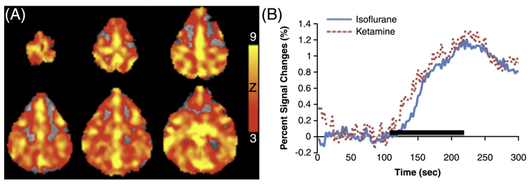
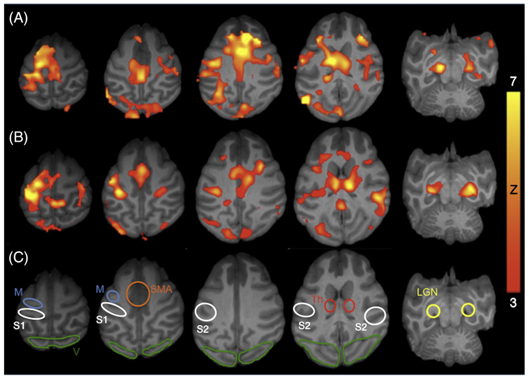
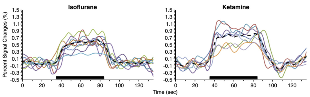
Baboon, with its large brain size and extensive cortical folding compared to other non-human primates, serves as a good model for neuroscience research. This study reports the implementation of a baboon model for blood oxygenation level-dependent (BOLD) fMRI studies (1.5 x 1.5 x 4 mm resolution) on a clinical 3T-MRI scanner. BOLD fMRI responses to hypercapnic (5% CO(2)) challenge, 10 Hz flicker visual, and vibrotactile somatosensory-motor stimulations were investigated in baboons anesthetized sequentially with isoflurane and ketamine. Hypercapnia evoked robust BOLD increases. Paralysis was determined to be necessary to achieve reproducible functional activations within and between subjects under our experimental conditions. With optimized anesthetic doses (0.8-1.0% isoflurane or 6-8 mg/kg/h ketamine) and adequate paralysis (vecuronium, 0.2 mg/kg), robust activations were detected in the visual (V), primary (S1) and secondary (S2) somatosensory, primary motor (M cortices), supplementary motor area (SMA), lateral geniculate nucleus (LGN) and thalamus (Th). Data were tabulated for 11 trials under isoflurane and 10 trials under ketamine on 5 baboons. S1, S2, M, and V activations were detected in essentially all trials (90-100% of the time, except 82% for S2 under isoflurane and 70% for M under ketamine). LGN activations were detected 64-70% of the time under both anesthetics. SMA and Th activations were detected 36-45% of the time under isoflurane and 60% of the time under ketamine. BOLD percent changes among different structures were slightly higher under ketamine than isoflurane (0.75% versus 0.58% averaging all structures), but none was statistically different (P>0.05). This baboon model offers an opportunity to non-invasively image brain functions and dysfunctions in large non-human primates.
Copyright 2010 Elsevier Inc. All rights reserved.
Figures

Similar articles
-
Baseline CBF, and BOLD, CBF, and CMRO2 fMRI of visual and vibrotactile stimulations in baboons.J Cereb Blood Flow Metab. 2011 Feb;31(2):715-24. doi: 10.1038/jcbfm.2010.154. Epub 2010 Sep 8. J Cereb Blood Flow Metab. 2011. PMID: 20827260 Free PMC article.
-
Mouse BOLD fMRI at ultrahigh field detects somatosensory networks including thalamic nuclei.Neuroimage. 2019 Jul 15;195:203-214. doi: 10.1016/j.neuroimage.2019.03.063. Epub 2019 Apr 1. Neuroimage. 2019. PMID: 30946950
-
Mouse fMRI under ketamine and xylazine anesthesia: Robust contralateral somatosensory cortex activation in response to forepaw stimulation.Neuroimage. 2018 Aug 15;177:30-44. doi: 10.1016/j.neuroimage.2018.04.062. Epub 2018 May 4. Neuroimage. 2018. PMID: 29730495
-
BOLD fMRI mapping of brain responses to nociceptive stimuli in rats under ketamine anesthesia.Med Eng Phys. 2008 Oct;30(8):953-8. doi: 10.1016/j.medengphy.2007.12.004. Epub 2008 Feb 20. Med Eng Phys. 2008. PMID: 18243035
-
BOLD fMRI and hemodynamic responses to somatosensory stimulation in anesthetized mice: spontaneous breathing vs. mechanical ventilation.NMR Biomed. 2020 Jul;33(7):e4311. doi: 10.1002/nbm.4311. Epub 2020 Apr 15. NMR Biomed. 2020. PMID: 32297409 Free PMC article.
Cited by
-
Repetitive Transcranial Magnetic Stimulation Educes Frequency-Specific Causal Relationships in the Motor Network.Brain Stimul. 2016 May-Jun;9(3):406-414. doi: 10.1016/j.brs.2016.02.006. Epub 2016 Feb 16. Brain Stimul. 2016. PMID: 26964725 Free PMC article.
-
Distinct BOLD fMRI Responses of Capsaicin-Induced Thermal Sensation Reveal Pain-Related Brain Activation in Nonhuman Primates.PLoS One. 2016 Jun 16;11(6):e0156805. doi: 10.1371/journal.pone.0156805. eCollection 2016. PLoS One. 2016. PMID: 27309348 Free PMC article.
-
Multimodal MRI of nonhuman primate stroke.Transl Stroke Res. 2012 Mar;3(1):84-9. doi: 10.1007/s12975-012-0145-1. Epub 2012 Feb 4. Transl Stroke Res. 2012. PMID: 24323756 Free PMC article.
-
MRI of perfusion-diffusion mismatch in non-human primate (baboon) stroke: a preliminary report.Open Neuroimag J. 2011;5:147-52. doi: 10.2174/1874440001105010147. Epub 2011 Nov 18. Open Neuroimag J. 2011. PMID: 22253656 Free PMC article.
-
Investigation of cross-species translatability of pharmacological MRI in awake nonhuman primate - a buprenorphine challenge study.PLoS One. 2014 Oct 22;9(10):e110432. doi: 10.1371/journal.pone.0110432. eCollection 2014. PLoS One. 2014. PMID: 25337714 Free PMC article.
References
-
- Andersen AH, Zhang Z, Barber T, Rayens WS, Zhang J, Grondin R, Hardy P, Gerhardt GA, Gash DM. Functional MRI studies in awake rhesus monkeys: methodological and analytical strategies. J. Neurosci. Methods. 2002;118:141–152. - PubMed
-
- Bianciardi M, Cerasa A, Patria F, Hagberg GE. Evaluation of mixed effects in event-related fMRI studies: impact of first-level design and filtering. NeuroImage. 2004;22:1351–1370. - PubMed
-
- Buxton RB, Uludağ K, Dubowitz DJ, Liu TT. Modeling the hemodynamic response to brain activation. NeuroImage. 2004;23 Suppl 1:S220–S233. - PubMed
-
- Chakravarty MM, Broadbent S, Rosa-Neto P, Lambert CM, Collins DL. Design, construction, and validation of an MRI-compatible vibrotactile stimulator intended for clinical use. J. Neurosci. Methods. 2009a;184:129–135. - PubMed
Publication types
MeSH terms
Grants and funding
LinkOut - more resources
Full Text Sources
Medical
