

Expression of leukemia/lymphoma-related factor (LRF/POKEMON) in human breast carcinoma and other cancers
- PMID: 20471975
- PMCID: PMC2939325
- DOI: 10.1016/j.yexmp.2010.05.002
Expression of leukemia/lymphoma-related factor (LRF/POKEMON) in human breast carcinoma and other cancers
Abstract
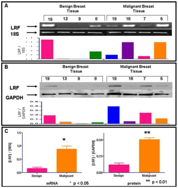
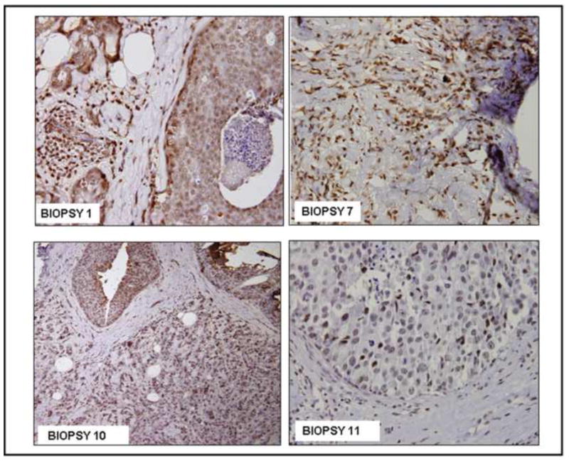
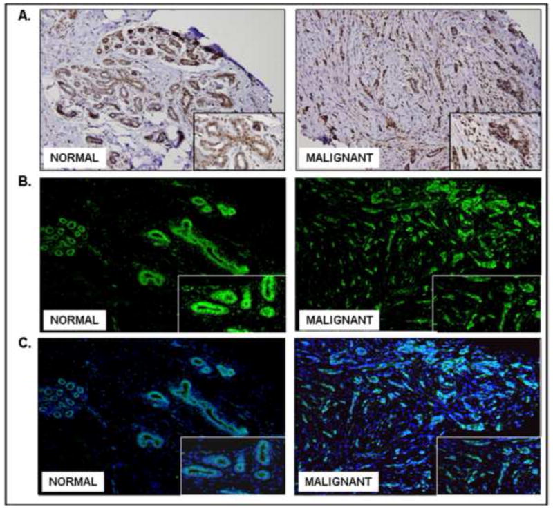
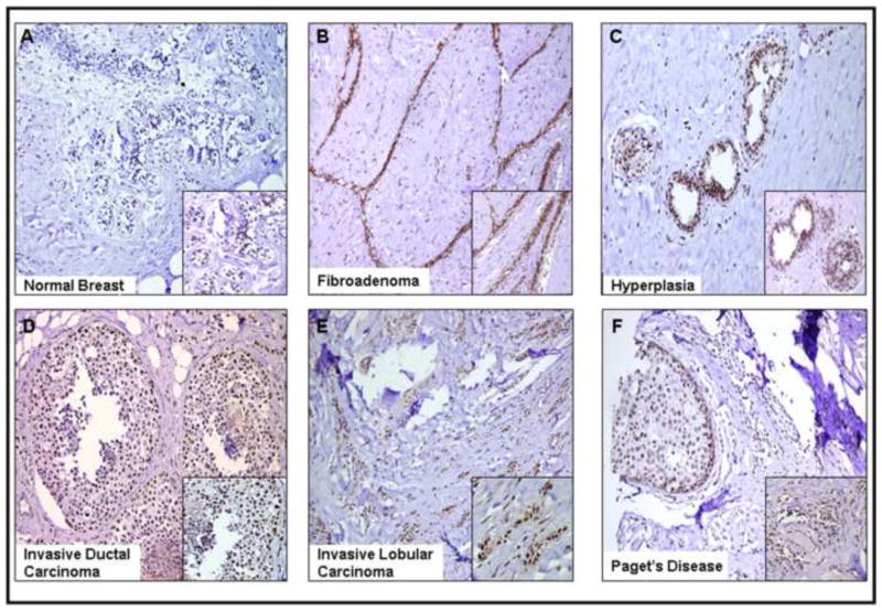
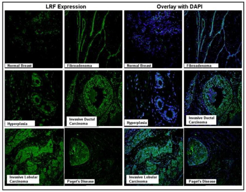
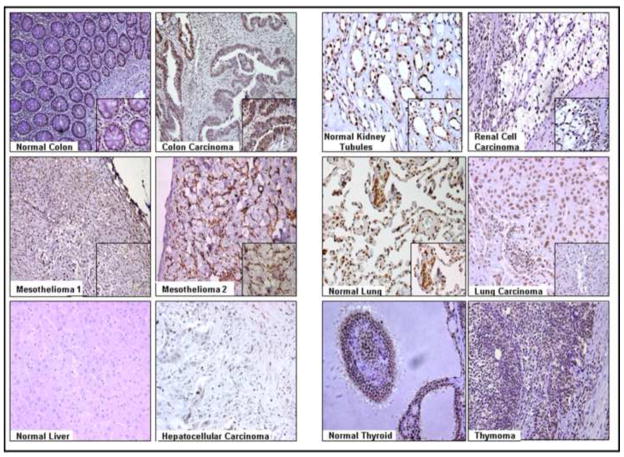
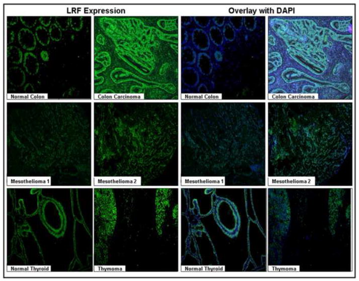
The POK family of proteins plays an important role in not only embryonic development and cell differentiation, but also in oncogenesis. Leukemia/lymphoma-related factor (LRF) belongs to the POK family of transcriptional repressors and is also known as POK erythroid myeloid ontogenic factor (POKEMON), which binds to short transcripts of HIV-1 (FBI-1) and TTF-1 interacting peptide (TIP21). Its oncogenic role is known only in lymphoma, non-small cell lung carcinoma, and malignant gliomas. The functional expression of LRF in human breast carcinoma has not yet been confirmed. The aim of this study was to investigate and compare the expression of LRF in human breast cancer tissues and other human tumors. The expression of LRF mRNA transcripts and protein was observed in twenty human benign and malignant breast biopsy tissues. Expression of LRF was observed in several formalin-fixed tissues by immunohistochemistry and immunofluorescence. All malignant breast tissues expressed mRNA transcripts and protein for LRF. However, 40% and 15% benign breast biopsy tissues expressed LRF mRNA transcripts and protein, respectively. The overall expression of LRF mRNA transcripts and total protein was significantly more in malignant breast tissues than the benign breast tissues. LRF expression was also observed in the nuclei of human colon, renal, lung, hepatocellular carcinomas and thymoma tumor cells. In general, a significantly higher expression of LRF was seen in malignant tissues than in the corresponding benign or normal tissue. Further studies are warranted to determine the malignant role of LRF in human breast carcinoma.
Copyright © 2010 Elsevier Inc. All rights reserved.
Figures
References
-
- Agrawal A, Yang J, Murphy RF, Agrawal DK. Regulation of the p14ARF-Mdm2-p53 pathway: an overview in breast cancer. Exp Mol Pathol. 2006;81:115–122. - PubMed
-
- Bos R, van Diest PJ, van der Groep P, Greijer AE, Hermsen MAJA, Heijnen I, Meijer GA, Baak JPA, Pinedo HM, Wall E, Shvarts A. Protein expression of B-cell lymphoma gene 6 (BCL-6) in invasive breast cancer is associated with cyclin D1 and hypoxia-inducible factor-1 alpha (HIF-1alpha) Oncogene. 2003;22:8948–8951. - PubMed
-
- Cui M, Xu H, Yang Y. Expression and clinical significance of Pokemon protein in breast cancer. J Clinical Surgery (Chinese) 2007;15:399–401.
-
- Davies JM, Hawe N, Kabaroswski J, Huan QH, Zhu J, Brand NJ, Leprince D, Dhordain P, Cook N, Morriss-Kay G, Zelenta A. Novel BTB/POZ domain zinc-finger protein, LRF, is a potential target of the LAZ-3/BCL-6 oncogene. Oncogene. 1999;18:365–375. - PubMed
Publication types
MeSH terms
Substances
Grants and funding
LinkOut - more resources
Full Text Sources
Medical
