Review
. 2010 Aug;87(2):162-78.
doi: 10.1016/j.antiviral.2010.05.002. Epub 2010 May 21.

Antiviral strategies to control calicivirus infections

Affiliations
Review

Antiviral strategies to control calicivirus infections

Jacques Rohayem et al. Antiviral Res. 2010 Aug.

Abstract

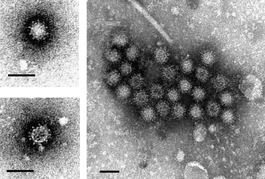
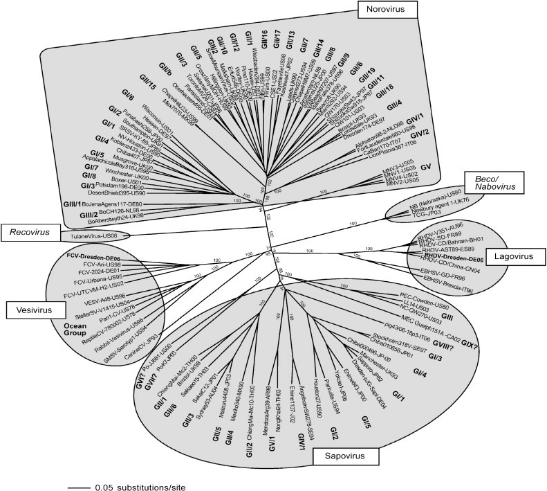
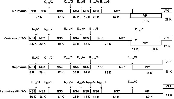
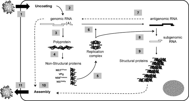
Caliciviridae are human or non-human pathogenic viruses with a high diversity. Some members of the Caliciviridae, i.e. human pathogenic norovirus or rabbit hemorrhagic disease virus (RHDV), are worldwide emerging pathogens. The norovirus is the major cause of viral gastroenteritis worldwide, accounting for about 85% of the outbreaks in Europe between 1995 and 2000. In the United States, 25 million cases of infection are reported each year. Since its emergence in 1984 as an agent of fatal hemorrhagic diseases in rabbits, RHDV has killed millions of rabbits and has been dispersed to all of the inhabitable continents. In view of their successful and apparently increasing emergence, the development of antiviral strategies to control infections due to these viral pathogens has now become an important issue in medicine and veterinary medicine. Antiviral strategies have to be based on an understanding of the epidemiology, transmission, clinical symptoms, viral replication and immunity to infection resulting from infection by these viruses. Here, we provide an overview of the mechanisms underlying calicivirus infection, focusing on the molecular aspects of replication in the host cell. Recent experimental data generated through an international collaboration on structural biology, virology and drug design within the European consortium VIZIER is also presented. Based on this analysis, we propose antiviral strategies that may significantly impact on the epidemiological characteristics of these highly successful viral pathogens.


Figures

Fig. 1
Fig. 1
Morphology of the human pathogenic norovirus. Clinical stool samples were negatively stained with 2% phosphotungstate, pH 7.0, and examined by transmission electron microscopy. The typical cup-like surface of the virions is visible. Scale bar = 50 nm.
Fig. 2
Fig. 2
Classification of the Caliciviridae. Phylogenetic analysis of strains belonging to the so far defined genera (Norovirus, Sapovirus, Lagovirus and Vesivirus) and the two tentative genera Beco/Nabovirus and Recovirus, as indicated. The complete capsid sequence of the strains was aligned with Clustal X (Gibson et al., 1999) and a phylogenetic analysis performed with the neighbour-joining method using PAUP (Swofford, 2002). A bootstrap analysis with 1000 replicates was performed. The bootstrap values are indicated. The strains belonging to non-defined genogroups (GVI. VII, VIII and IX of sapovirus) are indicated by a question mark. The strains FCV/Dresden/2006/GE strain (Vesivirus, GenBank accession No. DQ424892), and RHDV/Dresden/2006/GE (Lagovirus, GenBank accession no. EF363035) generated within the consortium VIZIER are highlighted in bold. The tentative Genera Recovirus and Beco/Nabovirus are indicated.
Fig. 3
Fig. 3
Comparison of the organization of the viral genome of the six calicivirus genera. The complete genome of the following strains is shown: Norovirus GII.4 (Hu/NLV/Dresden174/pUS-NorII/1997/GE strain, GenBank accession no. AY741811), vesivirus (feline calicivirus, FCV/Dresden/2006/GE strain, GenBank accession No. DQ424892), sapovirus GI.1 (Hu/SV/pJG-SapI/DE strain, GenBank accession no. AY694184), and lagovirus (RHDV strain RHDV/Dresden/2006/GE, GenBank accession no. EF363035). The cleavage sites in the viral polyprotein precursor are indicated, as well as the putative molecular weight in kDa (K) of the resulting non-structural proteins.
Fig. 4
Fig. 4
The multiplication cycle of the calicivirus. The replication of the calicivirus can be schematically subdivided into eleven steps. After attachment of the protein core to the cellular receptor, the virion is internalized in the cell (step 1). Uncoating of the viral genome (step 2) is followed by translation of the polyprotein precursor (step 3) and a co-translational processing releasing the non-structural proteins (step 4). Those proteins assemble in a replication complex (step 5) that synthesizes the antigenomic RNA (step 6), being itself used as a template for synthesis of the genomic RNA (step 7). The newly synthesized genomic RNA is translated as a polyprotein precursor (step 3) or being used for packaging in the assembled viral protein core (step 10). The antigenomic RNA is also the template for synthesis of subgenomic RNA (step 8). In noroviruses, vesiviruses and possibly recoviruses, the subgenomic RNA is translated as structural proteins, VP1 and VP2 (step 9). In sapoviruses, lagoviruses and possibly beco/naboviruses, the VP1 is released from the polyprotein precursor after processing by the viral protease. At a still not defined time in the multiplication cycle, assembly of the structural proteins as well as packaging of the genomic RNA occurs (step 10), followed by release of the mature virion from the cell (step 11).
Fig. 5
Fig. 5
Inhibition of calicivirus particle formation in CRFK-monolayer after infection with the FCV/Dresden/2006/GE strain (GenBank accession No. DQ424892). Two siRNA (siR19 and siR20) targeting conserved sequences in the 5′-UTR and 3′-UTR, respectively, were used. The siRNA (330 nM) was transfected into the CRFK cells 5 h prior to infection with FCV. Plaque assays were performed as described by others (Gray, 1999), yielding a viral titre of 500 PFU/ml. The siRNA completely inhibited viral multiplication in the CRFK cells.
Fig. 6
Fig. 6
Inhibition of the activity of norovirus and sapovirus RNA-dependent RNA polymerases by nucleoside analogues. (A) Assessment of the activity of the norovirus RNA-dependent RNA polymerase was determined as previously described (Rohayem et al., 2006a, Rohayem et al., 2006b). (B) Assessment of the activity of the sapovirus RNA-dependent RNA polymerase NS7pol was determined as previously described (Fullerton et al., 2007). 2′-Fluor-UTP, 2′-fluorouridine-5′-triphosphate. 2′-O-Methyl-UTP, 2′-O-methyluridine-5′-triphosphate. 2′Ara-UTP, 2′-arauridine-5′-triphosphate; 3′-deoxy-UTP; 3′-deoxyuridine-5′-triphosphate. 2′-O-Methyl-GTP, 2′-O,methylguanidine-5′-triphosphate. The mean and the standard error of the mean of three independent experiments are shown. CPM, counts per minute. Each of the compounds was incubated at a concentration of 50 μM.

References

    1. Allaire M., Chernaia M.M., Malcolm B.A., James M.N. Picornaviral 3C cysteine proteinases have a fold similar to chymotrypsin-like serine proteinases. Nature. 1994;369:72–76. - PubMed
    1. Anand K., Palm G.J., Mesters J.R., Siddell S.G., Ziebuhr J., Hilgenfeld R. Structure of coronavirus main proteinase reveals combination of a chymotrypsin fold with an extra alpha-helical domain. Embo J. 2002;21:3213–3224. - PMC - PubMed
    1. Anand K., Ziebuhr J., Wadhwani P., Mesters J.R., Hilgenfeld R. Coronavirus main proteinase (3CLpro) structure: basis for design of anti-SARS drugs. Science. 2003;300:1763–1767. - PubMed
    1. Anderson E.J. Prevention and treatment of viral diarrhea in pediatrics. Expert Rev. Anti Infect. Ther. 2010;8:205–217. - PubMed
    1. Asanaka M., Atmar R.L., Ruvolo V., Crawford S.E., Neill F.H., Estes M.K. Replication and packaging of Norwalk virus RNA in cultured mammalian cells. Proc. Natl. Acad. Sci. U.S.A. 2005 - PMC - PubMed

Publication types

MeSH terms