Current dipole orientation and distribution of epileptiform activity correlates with cortical thinning in left mesiotemporal epilepsy
- PMID: 20472073
- PMCID: PMC3043985
- DOI: 10.1016/j.neuroimage.2010.04.264
Current dipole orientation and distribution of epileptiform activity correlates with cortical thinning in left mesiotemporal epilepsy
Abstract
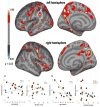
To evaluate cortical architecture in mesial temporal lobe epilepsy (MTLE) with respect to electrophysiology, we analyze both magnetic resonance imaging (MRI) and magnetoencephalography (MEG) in 19 patients with left MTLE. We divide the patients into two groups: 9 patients (Group A) have vertically oriented antero-medial equivalent current dipoles (ECDs). 10 patients (Group B) have ECDs that are diversely oriented and widely distributed. Group analysis of MRI data shows widespread cortical thinning in Group B compared with Group A, in the left hemisphere involving the cingulate, supramarginal, occipitotemporal and parahippocampal gyri, precuneus and parietal lobule, and in the right hemisphere involving the fronto-medial, -central and -basal gyri and the precuneus. These results suggest that regardless of the presence of hippocampal sclerosis, in a subgroup of patients with MTLE a large cortical network is affected. This finding may, in part, explain the unfavorable outcome in some MTLE patients after epilepsy surgery.
Copyright 2010 Elsevier Inc. All rights reserved.
Figures


Similar articles
-
High-resolution source imaging in mesiotemporal lobe epilepsy: a comparison between MEG and simultaneous EEG.J Clin Neurophysiol. 2003 Jul-Aug;20(4):227-38. doi: 10.1097/00004691-200307000-00001. J Clin Neurophysiol. 2003. PMID: 14530735
-
Functional substrate for memory function differences between patients with left and right mesial temporal lobe epilepsy associated with hippocampal sclerosis.Epilepsy Behav. 2015 Oct;51:251-8. doi: 10.1016/j.yebeh.2015.07.032. Epub 2015 Aug 24. Epilepsy Behav. 2015. PMID: 26300534
-
Mesial temporal lobe epilepsy with hippocampal sclerosis is a network disorder with altered cortical hubs.Epilepsia. 2015 May;56(5):772-9. doi: 10.1111/epi.12966. Epub 2015 Mar 23. Epilepsia. 2015. PMID: 25809843
-
Morphometric MRI features are associated with surgical outcome in mesial temporal lobe epilepsy with hippocampal sclerosis.Epilepsy Res. 2017 May;132:78-83. doi: 10.1016/j.eplepsyres.2017.02.022. Epub 2017 Mar 1. Epilepsy Res. 2017. PMID: 28324681
-
Subtypes of medial temporal lobe epilepsy: influence on temporal lobectomy outcomes?Epilepsia. 2012 Jan;53(1):1-6. doi: 10.1111/j.1528-1167.2011.03298.x. Epub 2011 Nov 2. Epilepsia. 2012. PMID: 22050314 Review.
Cited by
-
Clinical application of spatiotemporal distributed source analysis in presurgical evaluation of epilepsy.Front Hum Neurosci. 2014 Feb 10;8:62. doi: 10.3389/fnhum.2014.00062. eCollection 2014. Front Hum Neurosci. 2014. PMID: 24574999 Free PMC article. Review.
-
Multimodal Brain Network Jointly Construction and Fusion for Diagnosis of Epilepsy.Front Neurosci. 2021 Sep 29;15:734711. doi: 10.3389/fnins.2021.734711. eCollection 2021. Front Neurosci. 2021. PMID: 34658773 Free PMC article.
-
Analysis of Interictal Epileptiform Discharges in Mesial Temporal Lobe Epilepsy Using Quantitative EEG and Neuroimaging.Front Neurol. 2020 Nov 26;11:569943. doi: 10.3389/fneur.2020.569943. eCollection 2020. Front Neurol. 2020. PMID: 33324321 Free PMC article.
-
Altered structural connectome in temporal lobe epilepsy.Radiology. 2014 Mar;270(3):842-8. doi: 10.1148/radiol.13131044. Epub 2013 Nov 8. Radiology. 2014. PMID: 24475828 Free PMC article.
References
-
- Bernasconi N, Duchesne S, Janke A, Lerch J, Collins DL, Bernasconi A. Whole-brain voxel-based statistical analysis of gray matter and white matter in temporal lobe epilepsy. Neuroimage. 2004;23:717–723. - PubMed
-
- Dale AM, Fischl B, Sereno MI. Cortical surface-based analysis. I. Segmentation and surface reconstruction. Neuroimage. 1999;9:179–194. - PubMed
-
- Duzel E, Schiltz K, Solbach T, Peschel T, Baldeweg T, Kaufmann J, Szentkuti A, Heinze HJ. Hippocampal atrophy in temporal lobe epilepsy is correlated with limbic systems atrophy. J Neurol. 2006;253:294–300. - PubMed
Publication types
MeSH terms
Grants and funding
LinkOut - more resources
Full Text Sources
Medical

