Novel derivative of benzofuran induces cell death mostly by G2/M cell cycle arrest through p53-dependent pathway but partially by inhibition of NF-kappaB
- PMID: 20472557
- PMCID: PMC2903425
- DOI: 10.1074/jbc.M110.131797
Novel derivative of benzofuran induces cell death mostly by G2/M cell cycle arrest through p53-dependent pathway but partially by inhibition of NF-kappaB
Abstract
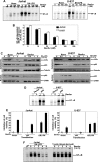
The Dracaena resin is widely used in traditional medicine as an anticancer agent, and benzofuran lignan is the active component. In this report, we provide evidence that the synthetic derivative of benzofuran lignan (Benfur) showed antitumor activities. It induced apoptosis in p53-positive cells. Though it inhibited endotoxin-induced nuclear factor kappaB (NF-kappaB) activation in both p53-positive and -negative cells, the activation of caspase 3 was observed in p53-positive cells. It showed partial cell death effect in both p53-positive and -negative cells through inhibition of NF-kappaB. Cell cycle analysis using flow cytometry showed that treatment with this novel benozofuran lignan derivative to Jurkat T-cells, but not U-937 cells, resulted in a G2/M arrest in a dose- and time-dependent manner. It increased amounts of p21, p27, and cyclin B, but not phospho-Rb through p53 nuclear translocation in Jurkat T-cells, but not in U-937 cells. It inhibited amounts of MDM2 (murine double minute 2) by repressing the transcription factor Sp1, which was also proved in silico. It induced cell death in tumor cells, but not in primary T-cells. Overall, our data suggest that Benfur-mediated cell death is partially dependent upon NF-kappaB, but predominantly dependent on p53. Thus, this novel benzofuran lignan derivative can be effective chemopreventive or chemotherapeutic agent against malignant T-cells.
Figures


Similar articles
-
Profilin potentiates chemotherapeutic agents mediated cell death via suppression of NF-κB and upregulation of p53.Apoptosis. 2016 Apr;21(4):502-13. doi: 10.1007/s10495-016-1222-9. Apoptosis. 2016. PMID: 26842845
-
Role of p53 and NF-kappaB in epigallocatechin-3-gallate-induced apoptosis of LNCaP cells.Oncogene. 2003 Jul 31;22(31):4851-9. doi: 10.1038/sj.onc.1206708. Oncogene. 2003. PMID: 12894226
-
Combretastatin-A4 prodrug induces mitotic catastrophe in chronic lymphocytic leukemia cell line independent of caspase activation and poly(ADP-ribose) polymerase cleavage.Clin Cancer Res. 2002 Aug;8(8):2735-41. Clin Cancer Res. 2002. PMID: 12171907
-
p53-independent roles of MDM2 in NF-κB signaling: implications for cancer therapy, wound healing, and autoimmune diseases.Neoplasia. 2012 Dec;14(12):1097-101. doi: 10.1593/neo.121534. Neoplasia. 2012. PMID: 23308042 Free PMC article. Review.
-
P53 vs NF-κB: the role of nuclear factor-kappa B in the regulation of p53 activity and vice versa.Cell Mol Life Sci. 2020 Nov;77(22):4449-4458. doi: 10.1007/s00018-020-03524-9. Epub 2020 Apr 22. Cell Mol Life Sci. 2020. PMID: 32322927 Free PMC article. Review.
Cited by
-
Development of novel benzofuran-isatin conjugates as potential antiproliferative agents with apoptosis inducing mechanism in Colon cancer.J Enzyme Inhib Med Chem. 2021 Dec;36(1):1424-1435. doi: 10.1080/14756366.2021.1944127. J Enzyme Inhib Med Chem. 2021. PMID: 34176414 Free PMC article.
-
Advanced glycation end products (AGEs) induce apoptosis via a novel pathway: involvement of Ca2+ mediated by interleukin-8 protein.J Biol Chem. 2011 Oct 7;286(40):34903-13. doi: 10.1074/jbc.M111.279190. Epub 2011 Aug 23. J Biol Chem. 2011. PMID: 21862577 Free PMC article.
-
P53 induction accompanying G2/M arrest upon knockdown of tumor suppressor HIC1 in U87MG glioma cells.Mol Cell Biochem. 2014 Oct;395(1-2):281-90. doi: 10.1007/s11010-014-2137-9. Epub 2014 Jul 4. Mol Cell Biochem. 2014. PMID: 24992983
-
Metallothionein 2A inhibits NF-κB pathway activation and predicts clinical outcome segregated with TNM stage in gastric cancer patients following radical resection.J Transl Med. 2013 Jul 19;11:173. doi: 10.1186/1479-5876-11-173. J Transl Med. 2013. PMID: 23870553 Free PMC article.
-
Cytotoxic compounds from the leaves and stems of the endemic Thai plant Mitrephora sirikitiae.Pharm Biol. 2020 Dec;58(1):490-497. doi: 10.1080/13880209.2020.1765813. Pharm Biol. 2020. PMID: 32478640 Free PMC article.
References
-
- Perez-Stable C. (2006) Cancer Lett. 231, 49–64 - PubMed
-
- Li Q., Sham H., Rosenberg S. (1999) Annual Reports in Medicinal Chemistry, Vol. 34, pp. 139–148, Academic Press Inc., New York
-
- Jordan A., Hadfield J. A., Lawrence N. J., McGown A. T. (1998) Med. Res. Rev. 18, 259–296 - PubMed
-
- Concin N., Stimpfl M., Zeillinger C., Wolff U., Hefler L., Sedlak J., Leodolter S., Zeillinger R. (2003) Int. J. Oncol. 22, 51–57 - PubMed
-
- Takizawa C. G., Morgan D. O. (2000) Curr. Opin. Cell Biol. 12, 658–665 - PubMed
Publication types
MeSH terms
Substances
LinkOut - more resources
Full Text Sources
Research Materials
Miscellaneous

